

Blaasjes uitgescheiden door bacteriën: een nieuw uitgangspunt kankeronderzoek?
Blaasjes uitgescheiden door bacteriën: een nieuw uitgangspunt voor kankeronderzoek?
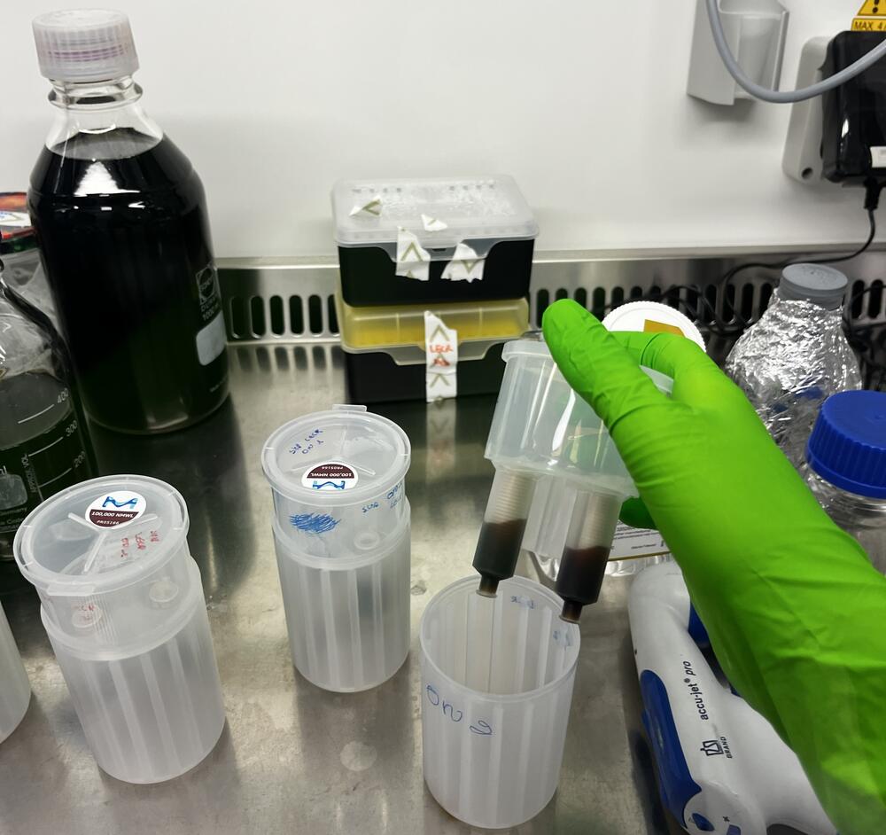
Stel je eens voor dat we blaasjes, van nature uitgescheiden door bacteriën, zouden kunnen inzetten als slimme medicijnen tegen kanker. Dat klinkt als verre toekomstmuziek, maar wereldwijd storten onderzoekers zich op dit interessante onderzoeksveld. Bacteriën scheiden deze blaasjes namelijk uit om onderling en met menselijke cellen te communiceren en kunnen zo invloed uitoefenen op ziekten zoals kanker. Om deze blaasjes ooit te kunnen gebruiken als kankermedicijn moeten we de blaasjes zuiver kunnen verzamelen en daar wringt het schoentje.
De rol van bacteriële blaasjes in onze gezondheid
De booschapperblaasjes, uitgescheiden door bacteriën in o.a. het menselijk lichaam, kunnen doorheen het lichaam reizen. Deze blaasjes dragen een lading met eiwitten, vetten en DNA/RNA van de bacterie als een soort USB-stick met instructies. Die lading kan alarmsensoren in ons afweersysteem activeren, waardoor ontstekingreacties of afweerreacties beïnvloed kunnen worden. In de masterproef van Shauni Van Rijckeghem, uitgevoerd in samenwerking met de onderzoekgroep Laboratory of Experimental Cancer Research (LECR), staat een goedaardige bacterie centraal. “Deze bacterie bevindt zich in de darmen en scheidt ook daar blaasjes uit. Die blaasjes kunnen door de darmwand worden opgenomen, om zo in de bloedbaan terecht te komen. Ze spelen mogelijk een belangrijke rol in verschillende ziekten, wat ze bijzonder interessant maakt om verder te ontwikkelen als therapie”, aldus Shauni.
Het obstakel: het kaf van het koren scheiden
Om bacteriële blaasjes goed te kunnen onderzoeken, stuiten onderzoekers op een praktisch probleem. Shauni legt uit dat de bacteriële blaasjes zich in het menselijk lichaam in een complexe omgeving bevinden, zoals bijvoorbeeld de darm of bloed. “Het is een uitdaging om de kwetsbare blaasjes zuiver en zonder ze te beschadigen uit deze complexe lichaamsvloeistoffen te isoleren. Dit komt omdat er in deze lichaamsvloeistoffen componenten zitten met gelijkaardige eigenschappen als bacteriële blaasjes.”
Je kunt het eigenlijk vergelijken met een zak vol knikkers, waarvan sommige dezelfde grootte hebben en anderen groter of kleiner zijn. Als je de knikkers zeeft, zullen sommige knikkers erdoor vallen en andere niet. Zo kan je al op een bepaalde manier een selectie maken, maar verschillende componenten met dezelfde grootte kan je zo onmogelijk van elkaar scheiden. Er zijn dus meerdere achtereenvolgende methodes nodig om de blaasjes van andere complexe componenten te kunnen scheiden.
Bovendien hebben bacteriën verschillende groeifasen. In de verschillende groeifasen scheiden bacteriën blaasjes met een andere samenstelling uit, wat ook een invloed kan hebben op hoe we de blaasjes moeten opzuiveren. Ze ‘groeien’ door zich in 2 te splitsen. Eerst passen ze zich aan hun omgeving. In deze fase splitsen en groeien ze nog niet. Nadat ze zich hebben aangepast aan hun omgeving, splitsen ze bacteriën en groeit elke individuele bacterie. Als de voedingsstoffen zijn opgebruikt, stoppen ze met groeien en sterven ze uiteindelijk af.
Een slimme dubbele scheidingsmethode
In haar onderzoek heeft Shauni twee scheidingsmethodes met elkaar vergeleken. Dit heeft ze gedaan zowel in de groeifase waarin de bacteriën sterk vermenigvuldigen (exponentiële groeifase) als in de groeifase waarin de bacteriën hun voedingsstoffen hebben opgebruikt en dus niet meer groeien (stationaire groeifase).
Omdat het scheiden van de blaasjes uit lichaamsvloeistoffen erg complex is, testte en controleerde ze de verschillende scheidingsmethoden in een kweekmedium in plaats van in lichaamsvloeistoffen. “De bacteriën werden eerst uit het medium verwijderd. Daarna wordt de overblijvende vloeistof gefilterd om grote componenten uit het medium te verwijderen. Om componenten verder van elkaar te scheiden wordt er een gradiënt met verschillende densiteiten gemaakt. Ze migreren dan naar de fracties met dezelfde densiteit.”
In het eerste experiment heeft ze dan alle componenten uit deze fracties verzameld. Uit een kleurentest, waarbij een blauwe kleur de aanwezigheid van blaasjes aantoont, bleek dat de blaasjes over meerdere fracties verspreid waren. “Dit kan wijzen op de aanwezigheid van andere componenten, die de test verstoren, in de opgezuiverde fracties”, aldus Shauni. “Daarom voegden we in het tweede experiment een extra scheidingsstap toe, waarbij we een methode gebruikten die de componenten in elke densiteitsfractie specifieker scheidt op basis van de grootte. Dan zagen we een duidelijke piek in een specifieke fractie, wat wijst op een succesvollere opzuivering van de blaasjes, gezien het gebrek aan storende signalen."


Kleurreactie die de aan- of afwezigheid van bacteriële blaasjes in verschillende densiteitsfracties aantoont. De donkerblauwe kleur in een specifieke densiteitsfractie, waargenomen voor beide groeifasen, laat zien dat de blaasjes met de gecombineerde methode gerichter en zuiverder verzameld zijn.
Toekomstperspectief
Het is cruciaal om de blaasjes zo goed mogelijk van de andere componenten te scheiden om het effect van de blaasjes betrouwbaar te kunnen onderzoeken. “We willen kijken hoe de blaasjes van deze specifieke bacterie het immuunsysteem beïnvloeden,” verduidelijkt Shauni. “Als er nog andere stoffen achterblijven, kunnen we nooit met zekerheid zeggen of het effect op het afweersysteem te wijten is aan de blaasjes zelf of aan andere stoffen die ook in het mengsel aanwezig zijn.”
Het is ook bekend dat bacteriële blaasjes een invloed kunnen hebben op de ontwikkeling van kanker. Sommige blaasjes, voornamelijk die afkomstig van goede bacteriën, kunnen afweercellen actiever maken en zo de afweer stimuleren om tumorgroei te remmen. Andere blaasjes, vaak afkomstig van ziekteverwekkers, kunnen dan weer de tumor helpen groeien door de afweer ‘in slaap te wiegen’. Doordat de afweer dan de tumor niet herkent, heeft de tumor de kans om verder te blijven groeien.
Bibliografie
REFERENCES
1. Xie J, Haesebrouck F, Van Hoecke L, Vandenbroucke RE. Bacterial extracellular vesicles: an emerging avenue to tackle diseases. Trends in Microbiology. Elsevier Ltd; 2023. p. 1206–24. doi:10.1016/j.tim.2023.05.010 PubMed PMID: 37330381.
2. Toyofuku M, Schild S, Kaparakis-Liaskos M, Eberl L. Composition and functions of bacterial membrane vesicles. Nature Reviews Microbiology. Nature Research; 2023. p. 415–30. doi:10.1038/s41579-023-00875-5 PubMed PMID: 36932221.
3. Welsh JA, Goberdhan DCI, O’Driscoll L, Buzas EI, Blenkiron C, Bussolati B, et al. Minimal information for studies of extracellular vesicles (MISEV2023): From basic to advanced approaches. J Extracell Vesicles. 2024 Feb 1;13(2). doi:10.1002/jev2.12404 PubMed PMID: 38326288.
4. Tulkens J, De Wever O, Hendrix A. Analyzing bacterial extracellular vesicles in human body fluids by orthogonal biophysical separation and biochemical characterization. Nat Protoc. 2020 Jan 1;15(1):40–67. doi:10.1038/s41596-019-0236-5 PubMed PMID: 31776460.
5. Brown L, Wolf JM, Prados-Rosales R, Casadevall A. Through the wall: Extracellular vesicles in Gram-positive bacteria, mycobacteria and fungi. Nature Reviews Microbiology. Nature Publishing Group; 2015. p. 620–30. doi:10.1038/nrmicro3480 PubMed PMID: 26324094.
6. Liu Y, Defourny KAY, Smid EJ, Abee T. Gram-positive bacterial extracellular vesicles and their impact on health and disease. Front Microbiol. 2018 Jul 9;9(JUL). doi:10.3389/fmicb.2018.01502 PubMed PMID: 30038605.
7. Hendrix A, De Wever O. Systemically circulating bacterial extracellular vesicles: origin, fate, and function. Trends in Microbiology. Elsevier Ltd; 2022. p. 213–6. doi:10.1016/j.tim.2021.12.012 PubMed PMID: 35033427.
8. Xu Y, Xie C, Liu Y, Qin X, Liu J. An update on our understanding of Gram-positive bacterial membrane vesicles: discovery, functions, and applications. Frontiers in Cellular and Infection Microbiology. Frontiers Media SA; 2023. doi:10.3389/fcimb.2023.1273813 PubMed PMID: 37860067.
9. Liang A, Korani L, Yeung CLS, Tey SK, Yam JWP. The emerging role of bacterial extracellular vesicles in human cancers. J Extracell Vesicles. 2024 Oct 8;13(10). doi:10.1002/jev2.12521
10. Popa SJ, Stewart SE. Socially Distanced Intercellular Communication: Mechanisms for Extracellular Vesicle Cargo Delivery. In. 2021. p. 179–209. doi:10.1007/978-3-030-67171-6_8
11. Orench-Rivera N, Kuehn MJ. Environmentally controlled bacterial vesicle-mediated export. Cellular Microbiology. Blackwell Publishing Ltd; 2016. p. 1525–36. doi:10.1111/cmi.12676 PubMed PMID: 27673272.
12. Mandal PK, Ballerin G, Nolan LM, Petty NK, Whitchurch CB. Bacteriophage infection of escherichia coli leads to the formation of membrane vesicles via both explosive cell lysis and membrane blebbing. Microbiology (United Kingdom). 2021;167(4). doi:10.1099/mic.0.001021 PubMed PMID: 33871329.
13. Kasman LM, Porter LD. Bacteriophages. 2026. PubMed PMID: 29630237.
14. Pérez-Cruz C, Carrión O, Delgado L, Martinez G, López-Iglesias C, Mercade E. New type of outer membrane vesicle produced by the gram-negative bacterium Shewanella vesiculosa M7T: Implications for DNA content. Appl Environ Microbiol. 2013 Mar;79(6):1874–81. doi:10.1128/AEM.03657-12 PubMed PMID: 23315742.
15. Turnbull L, Toyofuku M, Hynen AL, Kurosawa M, Pessi G, Petty NK, et al. Explosive cell lysis as a mechanism for the biogenesis of bacterial membrane vesicles and biofilms. Nat Commun. 2016 Apr 14;7. doi:10.1038/ncomms11220 PubMed PMID: 27075392.
16. Wang X, Thompson CD, Weidenmaier C, Lee JC. Release of Staphylococcus aureus extracellular vesicles and their application as a vaccine platform. Nat Commun. 2018 Dec 1;9(1). doi:10.1038/s41467-018-03847-z PubMed PMID: 29643357.
17. Abe K, Toyofuku M, Nomura N, Obana N. Autolysis-mediated membrane vesicle formation in Bacillus subtilis. Environ Microbiol. 2021 May 1;23(5):2632–47. doi:10.1111/1462-2920.15502 PubMed PMID: 33817925.
18. Andreoni F, Toyofuku M, Menzi C, Kalawong R, Shambat SM, François P, et al. Antibiotics Stimulate Formation of Vesicles in Staphylococcus aureus in both Phage-Dependent and-Independent Fashions and via Different Routes [Internet]. 2019. doi:10.1128/AAC
19. da Silva Barreira D, Lapaquette P, Ducassou JN, Couté Y, Guzzo J, Rieu A. Spontaneous Prophage Induction Contributes to the Production of Membrane Vesicles by the Gram-Positive Bacterium Lacticaseibacillus casei BL23. mBio. 2022 Sep 1;13(5). doi:10.1128/mbio.02375-22 PubMed PMID: 36200778.
20. Toyofuku M, Cárcamo-Oyarce G, Yamamoto T, Eisenstein F, Hsiao CC, Kurosawa M, et al. Prophage-triggered membrane vesicle formation through peptidoglycan damage in Bacillus subtilis. Nat Commun. 2017 Dec 1;8(1). doi:10.1038/s41467-017-00492-w PubMed PMID: 28883390.
21. Briaud P, Carroll RK. Extracellular vesicle biogenesis and functions in gram-positive bacteria. Infection and Immunity. American Society for Microbiology; 2020. doi:10.1128/IAI.00433-20 PubMed PMID: 32989035.
22. Bielaszewska M, Daniel O, Nyč O, Mellmann A. In vivo secretion of β-lactamase-carrying outer membrane vesicles as a mechanism of β-lactam therapy failure. Membranes (Basel). 2021 Nov 1;11(11). doi:10.3390/membranes11110806
23. Rumbo C, Fernández-Moreira E, Merino M, Poza M, Mendez JA, Soares NC, et al. Horizontal transfer of the OXA-24 carbapenemase gene via outer membrane vesicles: A new mechanism of dissemination of carbapenem resistance genes in Acinetobacter baumannii. Antimicrob Agents Chemother. 2011 Jul;55(7):3084–90. doi:10.1128/AAC.00929-10 PubMed PMID: 21518847.
24. Manning AJ, Kuehn MJ. Contribution of bacterial outer membrane vesicles to innate bacterial defense. BMC Microbiol. 2011;11. doi:10.1186/1471-2180-11-258 PubMed PMID: 22133164.
25. Kharina A, Podolich O, Faidiuk I, Zaika S, Haidak A, Kukharenko O, et al. Temperate bacteriophages collected by outer membrane vesicles in Komagataeibacter intermedius. J Basic Microbiol. 2015 Apr 1;55(4):509–13. doi:10.1002/jobm.201400711 PubMed PMID: 25589010.
26. McBroom AJ, Kuehn MJ. Release of outer membrane vesicles by Gram-negative bacteria is a novel envelope stress response. Mol Microbiol. 2007 Jan;63(2):545–58. doi:10.1111/j.1365-2958.2006.05522.x PubMed PMID: 17163978.
27. Wang M, Nie Y, Wu XL. Extracellular heme recycling and sharing across species by novel mycomembrane vesicles of a Gram-positive bacterium. ISME Journal. 2021 Feb 1;15(2):605–17. doi:10.1038/s41396-020-00800-1 PubMed PMID: 33037324.
28. Toyofuku M, Morinaga K, Hashimoto Y, Uhl J, Shimamura H, Inaba H, et al. Membrane vesicle-mediated bacterial communication. ISME Journal. 2017 Jun 1;11(6):1504–9. doi:10.1038/ismej.2017.13 PubMed PMID: 28282039.
29. Biller SJ, Lundeen RA, Hmelo LR, Becker KW, Arellano AA, Dooley K, et al. Prochlorococcus extracellular vesicles: molecular composition and adsorption to diverse microbes. Environ Microbiol. 2022 Jan 1;24(1):420–35. doi:10.1111/1462-2920.15834 PubMed PMID: 34766712.
30. Wang Y, Hoffmann JP, Chou CW, Höner zu Bentrup K, Fuselier JA, Bitoun JP, et al. Burkholderia thailandensis outer membrane vesicles exert antimicrobial activity against drug-resistant and competitor microbial species. Journal of Microbiology. 2020 Jul 1;58(7):550–62. doi:10.1007/s12275-020-0028-1 PubMed PMID: 32281050.
31. Yue H, Jiang J, Taylor AJ, Leite ADL, Dodds ED, Du L. Outer Membrane Vesicle-Mediated Codelivery of the Antifungal HSAF Metabolites and Lytic Polysaccharide Monooxygenase in the Predatory Lysobacter enzymogenes. ACS Chem Biol. 2021 Jun 18;16(6):1079–89. doi:10.1021/acschembio.1c00260 PubMed PMID: 34032403.
32. Afoshin AS, Kudryakova I V., Borovikova AO, Suzina NE, Toropygin IY, Shishkova NA, et al. Lytic potential of Lysobacter capsici VKM B-2533T: bacteriolytic enzymes and outer membrane vesicles. Sci Rep. 2020 Dec 1;10(1). doi:10.1038/s41598-020-67122-2 PubMed PMID: 32561806.
33. De Langhe N, Van Dorpe S, Guilbert N, Vander Cruyssen A, Roux Q, Deville S, et al. Mapping bacterial extracellular vesicle research: insights, best practices and knowledge gaps. Nature Communications . 2024 Dec 1;15(1). doi:10.1038/s41467-024-53279-1 PubMed PMID: 39482295.
34. Kaparakis-Liaskos M, Ferrero RL. Immune modulation by bacterial outer membrane vesicles. Nature Reviews Immunology. Nature Publishing Group; 2015. p. 375–87. doi:10.1038/nri3837 PubMed PMID: 25976515.
35. Li Y, Li J, Dullaart RPF, Moshage H. Bacterial Extracellular Vesicles in the Pathogenesis and Treatment of Liver Diseases. Journal of Gastroenterology and Hepatology (Australia). John Wiley and Sons Inc; 2026. doi:10.1111/jgh.70264
36. Lee KE, Kim JK, Han SK, Lee DY, Lee HJ, Yim SV, et al. The extracellular vesicle of gut microbial Paenalcaligenes hominis is a risk factor for vagus nerve-mediated cognitive impairment. Microbiome. 2020 Jul 15;8(1). doi:10.1186/s40168-020-00881-2 PubMed PMID: 32669127.
37. Chronopoulos A, Kalluri R. Emerging role of bacterial extracellular vesicles in cancer. Oncogene. Springer Nature; 2020. p. 6951–60. doi:10.1038/s41388-020-01509-3 PubMed PMID: 33060855.
38. Tulkens J, Vergauwen G, Van Deun J, Geeurickx E, Dhondt B, Lippens L, et al. Increased levels of systemic LPS-positive bacterial extracellular vesicles in patients with intestinal barrier dysfunction. Gut. BMJ Publishing Group; 2020. p. 191–3. doi:10.1136/gutjnl-2018-317726 PubMed PMID: 30518529.
39. Hendrix A, Lippens L, Pinheiro C, Théry C, Martin-Jaular L, Lötvall J, et al. Extracellular vesicle analysis. Nature Reviews Methods Primers. 2023 Jul 27;3(1):56. doi:10.1038/s43586-023-00240-z
40. Huang W, Shu C, Hua L, Zhao Y, Xie H, Qi J, et al. Modified bacterial outer membrane vesicles induce autoantibodies for tumor therapy. Acta Biomater. 2020 May 1;108:300–12. doi:10.1016/j.actbio.2020.03.030 PubMed PMID: 32251780.
41. Monguió-Tortajada M, Gálvez-Montón C, Bayes-Genis A, Roura S, Borràs FE. Extracellular vesicle isolation methods: rising impact of size-exclusion chromatography. Cellular and Molecular Life Sciences. Springer Science and Business Media Deutschland GmbH; 2019. p. 2369–82. doi:10.1007/s00018-019-03071-y PubMed PMID: 30891621.
42. Kusaka Y, Adachi Y. Determination of hydrodynamic diameter of small flocs by means of direct movie analysis of Brownian motion. Colloids Surf A Physicochem Eng Asp. 2007 Oct 1;306(1-3 SPEC. ISS.):166–70. doi:10.1016/j.colsurfa.2007.03.030
43. Kowkabany G, Bao Y. Nanoparticle Tracking Analysis: An Effective Tool to Characterize Extracellular Vesicles. Molecules. Multidisciplinary Digital Publishing Institute (MDPI); 2024. doi:10.3390/molecules29194672 PubMed PMID: 39407601.
44. Webber J, Clayton A. How pure are your vesicles? J Extracell Vesicles. 2013;2(1). doi:10.3402/jev.v2i0.19861
45. InvivoGen. HEK-Blue hTLR2 cells | Data sheet | InvivoGen [Internet]. Available from: https://www.invivogen.com/hek-blue-detection
46. Dinis JM, Barton DE, Ghadiri J, Surendar D, Reddy K, Velasquez F, et al. In search of an uncultured human-associated TM7 bacterium in the environment. PLoS One. 2011;6(6). doi:10.1371/journal.pone.0021280 PubMed PMID: 21701585.
47. Liu BD, Akbar R, Oliverio A, Thapa K, Wang X, Fan GC. BACTERIAL EXTRACELLULAR VESICLES IN THE REGULATION OF INFLAMMATORY RESPONSE AND HOST-MICROBE INTERACTIONS. Shock. Lippincott Williams and Wilkins; 2024. p. 175–88. doi:10.1097/SHK.0000000000002252 PubMed PMID: 37878470.
48. Doré E, Boilard E. Bacterial extracellular vesicles and their interplay with the immune system. Pharmacology and Therapeutics. Elsevier Inc.; 2023. doi:10.1016/j.pharmthera.2023.108443 PubMed PMID: 37210006.
49. Cicenia A, Santangelo F, Gambardella L, Pallotta L, Iebba V, Scirocco A, et al. Protective Role of Postbiotic Mediators Secreted by Lactobacillus rhamnosus GG Versus Lipopolysaccharide-induced Damage in Human Colonic Smooth Muscle Cells. J Clin Gastroenterol. 2016 Dec 1;50:S140–4. doi:10.1097/MCG.0000000000000681 PubMed PMID: 27741159.
50. Laakso K, Koskenniemi K, Koponen J, Kankainen M, Surakka A, Salusjärvi T, et al. Growth phase-associated changes in the proteome and transcriptome of Lactobacillus rhamnosus GG in industrial-type whey medium. Microb Biotechnol. 2011 Nov;4(6):746–66. doi:10.1111/j.1751-7915.2011.00275.x PubMed PMID: 21883975.
51. Shibatal KI, Fujita M, Morita M. Structure-function Relationship of Mycoplasmal Lipoproteins/lipopeptides and Their Recognition by Toll-like Receptor 2*. Oral Biosci. 2004.
52. Li CC, Hsu WF, Chiang PC, Kuo MC, Wo AM, Tseng YJ. Characterization of markers, functional properties, and microbiome composition in human gut-derived bacterial extracellular vesicles. Gut Microbes. 2023;15(2). doi:10.1080/19490976.2023.2288200 PubMed PMID: 38038385.
53. Nordin JZ, Lee Y, Vader P, Mäger I, Johansson HJ, Heusermann W, et al. Ultrafiltration with size-exclusion liquid chromatography for high yield isolation of extracellular vesicles preserving intact biophysical and functional properties. Nanomedicine. 2015 May 1;11(4):879–83. doi:10.1016/j.nano.2015.01.003 PubMed PMID: 25659648.
54. SEPHAROSE AND SEPHAROSE CL GEL FILTRATION MEDIA Exact replacement for Product Code 84963.
55. Villéger R, Saad N, Grenier K, Falourd X, Foucat L, Urdaci MC, et al. Characterization of lipoteichoic acid structures from three probiotic Bacillus strains: involvement of d-alanine in their biological activity. Antonie van Leeuwenhoek, International Journal of General and Molecular Microbiology. 2014 Oct 1;106(4):693–706. doi:10.1007/s10482-014-0239-8 PubMed PMID: 25090957.





