

Hoe smartphonefoto brandwondenzorg wereldwijd kan veranderen
Wereldwijd sterven jaarlijks 180 000 mensen aan de rechtstreekse gevolgen van brandwonden. De grootste oorzaak hiervoor is de ontbrekende toegang tot gespecialiseerde zorg op vele plaatsen in de wereld. Zelfs wanneer die zorg wel beschikbaar is, blijft een correcte diagnose van de brandwondendiepte moeilijk. Ik ontwikkelde daarom een AI-model dat op basis van een gewone foto de diepte van brandwonden kan inschatten.
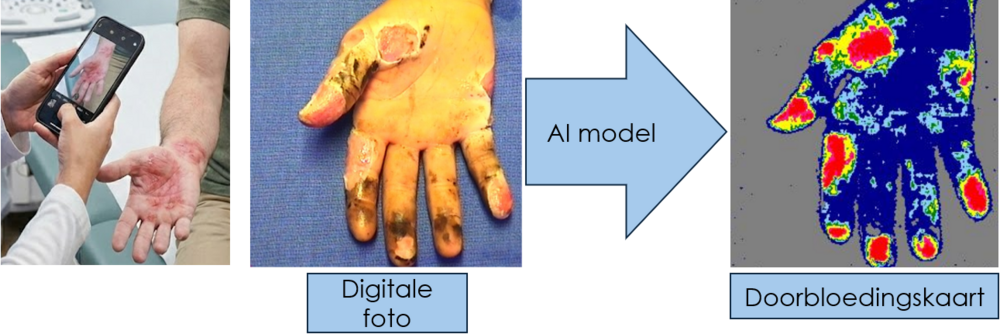
Van digitale foto naar doorbloedingskaart: het AI-model voorspelt op basis van een gewone foto (links) wat de dure laserscanner zou meten (rechts).
Een correcte inschatting van de diepte van brandwonden is cruciaal. Hier wordt bepaald of een wonde ofwel binnen 2-3 weken spontaan geneest, ofwel geopereerd moet worden om de dode huid te vervangen met een huidtransplantatie. Een onnodige operatie leidt tot een verhoogd risico op littekenvorming, langere ziekenhuisopnames en hogere kosten voor de maatschappij. Anderzijds, te laat opereren, verhoogt het risico op infecties en functionele beperkingen.
Kijken en gokken
 Vandaag de dag beoordeelt een arts brandwonden vooral met het blote oog. Hierbij wordt gefocust op de al dan niet aanwezigheid van blaren, de kleur en de pijngevoeligheid van de brandwonde. De techniek is snel, maar niet altijd even betrouwbaar. In 4 op 10 gevallen zitten zelfs de meest ervaren artsen naast de correcte diagnose.
Als alternatief bestaat er een laserscanner die de bloeddoorstroming in de brandwonde meet en in kaart brengt. Dit apparaat is veel nauwkeuriger om de branddiepte te meten, alleen kost zo’n toestel meer dan €70 000, en kost het onderhoud ervan jaarlijks €9 000. Bovendien is het apparaat niet draagbaar, neemt het veel tijd in beslag om een meting uit te voeren terwijl de patiënt helemaal stil moet blijven liggen, en is het gebruik bij jonge kinderen of zwaargewonde patiënten vaak geen optie. Daarom is deze scanner enkel te vinden in gespecialiseerde brandwondencentra met voldoende middelen.
Van foto naar diagnose
Net dat gebrek aan toegankelijkheid vormde voor mij de aanleiding om een vernieuwde techniek te ontwikkelen die goedkoop én nauwkeurig is. Hiervoor trainde ik een AI-model op duizenden foto’s en corresponderende laserscans van echte patiënten, met als doel om op basis van enkel een digitale foto van de brandwonde te voorspellen wat de accurate laserscanner zou meten.
Een goede diagnose van een brandwonde mag niet afhangen of een ziekenhuis een toestel van €70 000 kan betalen.
Het idee hiervoor is niet nieuw. Eerder toonde een onderzoek van de Université Libre de Bruxelles (ULB) al aan dat het mogelijk is om de diepte van een brandwonde af te leiden uit een gewone foto. Alleen vroeg deze methode wel handmatig bewerkte foto’s en negeerde het informatie over de patiënt zelf. Met die werkpunten ging ik aan de slag. Mijn model verwerkt de beelden automatisch en combineert de foto met cruciale klinische gegevens, zoals de leeftijd van de patiënt en het type verbranding. Een oudere huid reageert immers helemaal anders op hitte dan een jonge huid. Dankzij die extra gegevens te combineren met de foto, voorspelt het algoritme de bloeddoorstroming een stuk nauwkeuriger. Vergeleken met de eerdere AI-poging van de ULB, ligt de voorspelling van het vernieuwde model gemiddeld een vijfde dichter bij de werkelijke laserscans, en komen de voorspelde doorbloedingszones 80% beter overeen met de werkelijkheid.
Van ramp tot huisarts tot ontwikkelingsland
 De potentiële impact van deze nieuwe aanpak kan groot zijn. Denk aan rampen met veel slachtoffers, zoals de brand in Crans-Montana tijdens de nieuwjaarsnacht van 2025 naar 2026, waarbij 41 doden en 115 gewonden vielen. In zo’n noodsituatie is een laserscanner onbruikbaar. Toch moeten hulpverleners ter plaatse snel beslissen wie naar een gespecialiseerd brandwondencentrum moet en wie naar een plaatselijk ziekenhuis kan. Met een snelle smartphonefoto zou het nieuwe AI-model hen binnen enkele seconden kunnen helpen bij die belangrijke triage. Zo kunnen zwaargewonden sneller de juiste zorg krijgen en raken de gespecialiseerde brandwondencentra minder snel overspoeld.
Ook bij huisartsen kan de techniek een eerste, objectieve inschatting geven van de ernst van de brandwonde. Lichte brandwonden kunnen ter plaatse behandeld worden, terwijl ernstigere wonden worden doorverwezen. Dit ontlast brandwondencentra, die zo meer tijd en middelen overhouden voor wie er echt nood aan heeft. Toch blijft het model een hulpmiddel, want de arts blijft de diagnose stellen en de behandeling bepalen. Het gaat niet er niet om de arts te vervangen, maar om deze een extra ondersteuning bieden, zeker waar er geen laserscanners beschikbaar zijn.
Het gaat er niet om de arts te vervangen. Het model moet extra ondersteuning bieden, zeker waar er geen laserscanner is.
 Dat laatste geldt vooral voor ziekenhuizen en regio’s zonder het budget voor dure apparatuur. Het AI-model vereist enkel een digitale camera, zoals een smartphone, voor het nemen van de foto. Die is vele malen goedkoper dan een laserscanner van €70 000, die bovendien jaarlijks €9 000 aan onderhoud kost. Zo kunnen ook de kleinere ziekenhuizen en ontwikkelingslanden in de toekomst betrouwbare inschattingen maken, in plaats van volledig te moeten steunen op de expertise van de dokter, wat vandaag tot meer foute diagnoses leidt. Op die manier draagt het project rechtstreeks bij aan een duurzame gezondheidszorg, waarin kwalitatieve zorg voor iedereen betaalbaar en toegankelijk wordt.
Nog niet onmiddellijk in de app store
Hoewel de eerste resultaten veelbelovend lijken, zal de software nog niet onmiddellijk beschikbaar zijn in de app store. Zo moet er eerst nog samenwerkingen worden aangegaan met andere internationale brandwondencentra om het systeem te testen op verschillende cameratypes en huidtinten. Pas daarna kan de technologie veilig de stap maken van het lab naar de dagelijkse praktijk.
De eerste resultaten zijn veelbelovend, maar eerst moet er nog getest worden op verschillende cameratypes en huidtinten voor het in de app store kan.
Dit project toont wel al aan dat objectieve brandwondendiagnose niet onbetaalbaar hoeft te zijn. Door AI te combineren met een gewone foto, wil ik die diagnose toegankelijk maken voor iedereen, overal ter wereld. Op deze manier moet de arts geen toestel van €70 000 kopen, maar kan hij gebruik maken van iets dat hij al in zijn broekzak heeft.
Bibliografie
[1] World Health Organization. Burns. Oct. 2023. url: https://www.who.int/ news-room/fact-sheets/detail/burns (visited on 11/04/2025).
[2] Stichting Brandwonden. Epidemiologie. url: https://www.brandwonden. be/nl/leven-met-brandwonden/eerste-hulp/epidemiologie (visited on 02/02/2026).
[3] Monstrey, S. et al. “Assessment of burn depth and burn wound healing poten tial”. In: Burns 34.6 (2008), pp. 761–769. doi: 10.1016/j.burns.2008.01. 009.
[4] National Institute for Health and Care Excellence. MoorLDI2-BI: a laser Doppler blood flow imager for burn wound assessment. Medtech innovation briefing [MIB101]. Tech. rep. NICE, 2017. url: https://www.nice.org.uk/ guidance/mib101.
[5] LeCun, Y., Bengio, Y., and Hinton, G. “Deep learning”. In: Nature 521.7553 (2015), pp. 436–444. doi: 10.1038/nature14539.
[6] Carion, N. et al. SAM 3: Segment anything with concepts. 2025. doi: 10. 48550/arXiv.2511.16719.
[7] Siméoni, O. et al. DINOv3. arXiv. 2025. doi: 10.48550/arXiv.2508.10104.
[8] Perez, E. et al. FiLM: Visual reasoning with a general conditioning layer. arXiv. 2017. doi: 10.48550/arXiv.1709.07871.
[9] Cao, W., Mirjalili, V., and Raschka, S. “Rank consistent ordinal regression for neural networks with application to age estimation”. In: Pattern Recognition Letters 140 (2020), pp. 325–331. doi: 10.1016/j.patrec.2020.11.008.
[10] Shpichka, A. et al. “Skin tissue regeneration for burn injury”. In: Stem Cell Research & Therapy 10 (2019). doi: 10.1186/s13287-019-1203-3.
[11] Brownson, E. G. and Gibran, N. S. “Evaluation of the Burn Wound: Manage ment Decisions”. In: Total Burn Care. Ed. by D. N. Herndon. 5th ed. Elsevier, 2018, pp. 87–92. doi: 10.1016/B978-0-323-47661-4.00010-1.
[12] Cracowski, J.-L. and Roustit, M. “Human Skin Microcirculation”. In: Com prehensive Physiology 10.3 (2020), pp. 1105–1154. doi: 10.1002/j.2040 4603.2020.tb00131.x.
[13] Lee, Y. and Hwang, K. “Skin thickness of Korean adults”. In: Surgical and Radiologic Anatomy 24 (2002), pp. 183–189. doi: 10.1007/s00276-002 0034-5.
[14] DeSanti, L. “Pathophysiology and current management of burn injury”. In: Advances in Skin & Wound Care 18.6 (2005), pp. 323–332. doi: 10.1097/ 00129334-200507000-00013.
[15] Enoch, S., Roshan, A., and Shah, M. “Emergency and early management of burns and scalds”. In: BMJ 338 (2009), b1037.
[16] Warby, R. and Maani, C. V. “Burn classification”. In: StatPearls. StatPearls Publishing, 2023. url: https://www.ncbi.nlm.nih.gov/books/NBK539773/.
[17] Milner, S. M. “Classification of Burn Depth”. In: Eplasty 24 (2024), QA5.
[18] Jackson, D. M. “The diagnosis of the depth of burning”. In: British Journal of Surgery 40.164 (1953), pp. 588–596. doi: 10.1002/bjs.18004016413.
[19] Hettiaratchy, S. “Pathophysiology and types of burns”. In: BMJ 328.7453 (2004), pp. 1427–1429. doi: 10.1136/bmj.328.7453.1427.
[20] Li, H. et al. “Non-invasive medical imaging technology for the diagnosis of burn depth”. In: International Wound Journal 21 (2024). doi: 10.1111/iwj.14681.
[21] Stiles. Burn wound progression and the importance of first aid. https:// wounds-uk.com/journal-articles/burn-wound-progression-and-the importance-of-first-aid/. July 2015.
[22] Church, D. et al. “Burn Wound Infections”. In: Clinical Microbiology Reviews 19 (2006), pp. 403–434. doi: 10.1128/cmr.19.2.403-434.2006.
[23] Hermans, M. H. “A general overview of burn care”. In: International Wound Journal 2 (2005), pp. 206–220. doi: 10.1111/j.1742-4801.2005.00129.x.
[24] Loller, C., Buxton, G. A., and Kerzmann, T. L. “Hot soup! Correlating the severity of liquid scald burns to fluid and biomedical properties”. In: Burns 42.3 (May 2016), pp. 589–597. doi: 10.1016/j.burns.2015.10.016.
[25] Helanterä, I. et al. “The risk for end-stage renal disease is increased after burn”. In: Burns 42.2 (2016), pp. 316–321. issn: 0305-4179. doi: 10.1016/j.burns. 2015.10.027.
[26] Bounds, E. J. et al. “Electrical burns”. In: StatPearls. StatPearls Publishing, 2023. url: https://www.ncbi.nlm.nih.gov/books/NBK519514/.
[27] Heil, K. et al. “Freezing and non-freezing cold weather injuries: a systematic review”. In: British Medical Bulletin 117.1 (2016), pp. 79–93. doi: 10.1093/ bmb/ldw001.
[28] Zaramo, T. Z., Green, J. K., and Janis, J. E. “Practical Review of the Current Management of Frostbite Injuries”. In: Plastic and Reconstructive Surgery Global Open 10.10 (2022), e4618. doi: 10.1097/GOX.0000000000004618.
[29] Devgan, L. et al. “Modalities for the Assessment of Burn Wound Depth”. In: Journal of Burns and Wounds 5 (2006), e2. url: https://www.ncbi.nlm. nih.gov/pmc/articles/PMC1687143/.
[30] Monstrey, S. et al. “Assessment of burn depth and burn wound healing poten tial”. In: Burns 34.6 (2008), pp. 761–769. doi: 10.1016/j.burns.2008.01.009.
[31] Shin, H.-C. et al. “Deep Convolutional Neural Networks for Computer-Aided Detection: CNN Architectures, Dataset Characteristics and Transfer Learn ing”. In: IEEE Transactions on Medical Imaging 35 (2016), pp. 1285–1298. doi: 10.1109/tmi.2016.2528162.
[32] Jaskille, A. D. et al. “Critical Review of Burn Depth Assessment Techniques: Part II. Review of Laser Doppler Technology”. In: Journal of Burn Care & Research 31.1 (2010), pp. 151–157. doi: 10.1097/BCR.0b013e3181c7ed60.
[33] Khatib, M. et al. “A Systematic Review of the Evolution of Laser Doppler Techniques in Burn Depth Assessment”. In: Plastic Surgery International 2014 (2014), p. 621792. doi: 10.1155/2014/621792.
[34] Rajan, V. et al. “Review of methodological developments in laser Doppler flowmetry”. In: Lasers in Medical Science 24.2 (2009), pp. 269–283. doi: 10. 1007/s10103-007-0524-0.
[35] Lookeren Campagne, M. J. A. van. “Laser Doppler Flowmetry, using in vitro phantoms to study skin microcirculation”. Bachelor thesis. University of Twente, 2023. url: https://purl.utwente.nl/essays/94921.
[36] Moor Instruments. Basic Theory and Operating Principles of Laser Doppler Blood Flow Monitoring and Imaging (LDF & LDI). Tech. rep. Issue 1. Moor Instruments, 2018.
[37] A. P. Shepherd and P. A. Oberg, eds. Laser-Doppler Blood Flowmetry. Boston, MA: Springer Science+Business Media, 1990. doi: 10.1007/978-1-4757 2083-9.
[38] Fredriksson, I. et al. “Improved calibration procedure for laser Doppler perfu sion monitors”. In: Advanced Biomedical and Clinical Diagnostic Systems IX. Vol. 7890. SPIE, 2011, 78900G. doi: 10.1117/12.871938.
[39] Pape, S. A. et al. “Burn wound healing time assessed by laser Doppler imaging (LDI). Part 1: Derivation of a dedicated colour code for image interpretation”. In: Burns 38.2 (2012), pp. 187–194. doi: 10.1016/j.burns.2010.08.013.
[40] Droog, E. J., Steenbergen, W., and Sjoberg, F. “Measurement of depth of burns by laser Doppler perfusion imaging”. In: Burns 27.6 (2001), pp. 561 568. doi: 10.1016/S0305-4179(01)00021-3.
[41] Gill, P. “The critical evaluation of laser Doppler imaging in determining burn depth”. In: International Journal of Burn Trauma 3.2 (2013), pp. 72–77. url: https://www.ncbi.nlm.nih.gov/pmc/articles/PMC3636664/.
[42] Wang, R. et al. “Diagnostic Accuracy of Laser Doppler Imaging for the Assess ment of Burn Depth: A Meta-analysis and Systematic Review”. In: Journal of Burn Care & Research 41.3 (May 2020), pp. 619–625. doi: 10.1093/jbcr/ irz203.
[43] Moura et al. “Artificial intelligence in the management and treatment of burns: a systematic review”. In: Burns & Trauma 9 (2021).
[44] Taib et al. “Artificial intelligence in the management and treatment of burns: A systematic review and meta-analyses”. In: Journal of Plastic, Reconstructive & Aesthetic Surgery 77 (2023).
[45] Litjens, G. et al. “A survey on deep learning in medical image analysis”. In: Medical Image Analysis 42 (2017), pp. 60–88. doi: 10.1016/j.media.2017. 07.005.
[46] Ronneberger, O., Fischer, P., and Brox, T. “U-Net: Convolutional networks for biomedical image segmentation”. In: Lecture Notes in Computer Science. 2015, pp. 234–241. doi: 10.1007/978-3-319-24574-4\_28.
[47] Zhou, Z. et al. “UNet++: A nested UNet architecture for medical image seg mentation”. In: Deep Learning in Medical Image Analysis and Multimodal Learning for Clinical Decision Support. 2018, pp. 3–11. doi: 10.1007/978-3 030-00889-5\_1.
[48] Oktay, O. et al. “Attention U-Net: Learning where to look for the pancreas”. In: Medical Imaging with Deep Learning (2018). doi: 10.48550/arXiv.1804. 03999.
[49] Dosovitskiy, A. et al. “An image is worth 16x16 words: Transformers for image recognition at scale”. In: International Conference on Learning Representa tions. 2021. doi: 10.48550/arXiv.2010.11929.
[50] Vaswani, A. et al. “Attention is all you need”. In: Advances in Neural In formation Processing Systems. Vol. 30. 2017, pp. 5998–6008. url: https:// proceedings.neurips.cc/paper/2017/hash/3f5ee243547dee91fbd053c1c4a845aa Abstract.html.
[51] Simonyan, K. and Zisserman, A. “Very deep convolutional networks for large scale image recognition”. In: International Conference on Learning Represen tations. 2015. doi: 10.48550/arXiv.1409.1556.
[52] Rozo, A. et al. “A Deep Learning Image-to-Image Translation Approach for a More Accessible Estimator of the Healing Time of Burns”. In: IEEE Trans actions on Biomedical Engineering 70 (2023), pp. 2886–2894. doi: 10.1109/ tbme.2023.3267600.
[53] Bommasani, R. et al. On the opportunities and risks of foundation models. arXiv. 2021. doi: 10.48550/arXiv.2108.07258.
[54] Caron, M. et al. “Emerging properties in self-supervised vision transform ers”. In: Proceedings of the IEEE/CVF International Conference on Com puter Vision. 2021, pp. 9650–9660. url: https://openaccess.thecvf. com/content/ICCV2021/html/Caron_Emerging_Properties_in_Self Supervised_Vision_Transformers_ICCV_2021_paper.html.
[55] Liu, Z. et al. “A ConvNet for the 2020s”. In: Proceedings of the IEEE/CVF Conference on Computer Vision and Pattern Recognition. 2022, pp. 11976 11986. url: https://openaccess.thecvf.com/content/CVPR2022/html/ Liu_A_ConvNet_for_the_2020s_CVPR_2022_paper.html.
[56] Lemay, A. et al. “Benefits of linear conditioning with metadata for image segmentation”. In: Proceedings of the 4th Conference on Medical Imaging with Deep Learning. Vol. 143. 2021, pp. 396–414. url: https://proceedings. mlr.press/v143/lemay21a.html.
[57] Gutiérrez, P. A. et al. “Ordinal Regression Methods: Survey and Experimental Study”. In: IEEE Transactions on Knowledge and Data Engineering 28 (2015). doi: 10.1109/TKDE.2015.2457911.
[58] Selvaraju, R. R. et al. “Grad-CAM: Visual Explanations from Deep Networks via Gradient-Based Localization”. In: International Journal of Computer Vi sion 127.3 (2019), pp. 232–259. doi: 10.1007/s11263-019-01228-7.
[59] Gal, Y. and Ghahramani, Z. “Dropout as a Bayesian Approximation: Repre senting Model Uncertainty in Deep Learning”. In: Proceedings of the 33rd In ternational Conference on Machine Learning (ICML). PMLR. 2016, pp. 1050 1059. doi: 10.5555/3045390.3045502.
[60] Leibig, C. et al. “Leveraging uncertainty information from deep neural net works for disease detection”. In: Scientific Reports 7.1 (2017), p. 17816. doi: 10.1038/s41598-017-17876-z.
[61] Jadon, S. “A survey of loss functions for semantic segmentation”. In: (2020), pp. 1–7. doi: 10.1109/CIBCB48159.2020.9277638.
[62] Yeung, M. et al. “Unified Focal loss: Generalising Dice and cross entropy-based losses to handle class imbalanced medical image segmentation”. In: Comput erized Medical Imaging and Graphics 95 (2022), p. 102026. doi: 10.1016/j. compmedimag.2021.102026.
[63] Lin, T.-Y. et al. “Focal loss for dense object detection”. In: (2017), pp. 2980 2988. doi: 10.1109/ICCV.2017.324.
[64] Mathieu, M., Couprie, C., and LeCun, Y. “Deep multi-scale video prediction beyond mean square error”. In: (2016). doi: 10.48550/arXiv.1511.05440.
[65] Shan, G. and Jia, S. Convolution-based Probability Gradient Loss for Semantic Segmentation. 2024. doi: 10.48550/arXiv.2404.06704. url: https:// arxiv.org/abs/2404.06704.
[66] Kendall, A., Gal, Y., and Cipolla, R. “Multi-task learning using uncertainty to weigh losses for scene geometry and semantics”. In: Proceedings of the IEEE Conference on Computer Vision and Pattern Recognition (CVPR). 2018, pp. 7482 7491. doi: 10.1109/CVPR.2018.00781.
[67] Lakkapragada, A. et al. “Mitigating Negative Transfer in Multi-Task Learning with Exponential Moving Average Loss Weighting Strategies”. In: 37.13 (2023), pp. 16254–16255. doi: 10.1609/aaai.v37i13.26941.
[68] Wang, Z. et al. “Image quality assessment: from error visibility to structural similarity”. In: IEEE Transactions on Image Processing 13.4 (2004), pp. 600 612. doi: 10.1109/TIP.2003.819861.
[69] Frye, K. et al. “Simulated biologic intelligence used to predict length of stay and survival of burns”. In: Journal of Burn Care & Rehabilitation 17.6 (1996), pp. 540–546. doi: 10.1097/00004630-199611000-00011.
[70] Acha, B. et al. “CAD tool for burn diagnosis”. In: Information Processing in Medical Imaging. 2003, pp. 294–305. doi: 10.1007/978-3-540-45087-0\_25.
[71] Serrano, C. et al. “Features identification for automatic burn classification”. In: Burns 41.8 (2015), pp. 1883–1890. doi: 10.1016/j.burns.2015.05.011.
[72] Lee, J. et al. “Comparing Artificial Intelligence Guided Image Assessment to Current Methods of Burn Assessment”. In: Journal of Burn Care & Research 46.1 (2025), pp. 6–13. doi: 10.1093/jbcr/irae121.
[73] Abubakar, A., Ugail, H., Smith, K. M., et al. “Burns Depth Assessment Using Deep Learning Features”. In: Journal of Medical and Biological Engineering 40.6 (2020), pp. 923–933. doi: 10.1007/s40846-020-00574-z. url: https: //doi.org/10.1007/s40846-020-00574-z.
[74] Liu, H. et al. “A framework for automatic burn image segmentation and burn depth diagnosis using deep learning”. In: Computational and Mathematical Methods in Medicine 2021 (2021), pp. 1–12.
[75] Jiao, C. et al. “Burn image segmentation based on Mask Regions with Con volutional Neural Network deep learning framework: more accurate and more convenient”. In: Burns & Trauma 7 (2019), p. 6. doi: 10.1186/s41038-018 0137-9.
[76] Şevik, U. et al. “Automatic classification of skin burn colour images using texture-based feature extraction”. In: IET Image Processing 13 (2019), pp. 2018 2028.
[77] Chang, C. et al. “Deep learning-assisted burn wound diagnosis: Diagnostic model development study”. In: JMIR Medical Informatics 9 (2021), e22798.
[78] Thatcher, J. et al. “Imaging Techniques for Clinical Burn Assessment with a Focus on Multispectral Imaging”. In: Advances in Wound Care 5.8 (2016), pp. 360–378. doi: 10.1089/wound.2015.0684.
[79] Burmeister, D. et al. “Noninvasive Techniques for the Determination of Burn Severity in Real Time”. In: Journal of Burn Care & Research 38 (2017), e180 e191. doi: 10.1097/bcr.0000000000000338.
[80] Chen, J. et al. “Medical image translation with deep learning: Advances, datasets and perspectives”. In: Medical Image Analysis 103 (2025), p. 103605. doi: 10.1016/j.media.2025.103605.
[81] MoorInstruments. Single-Point Laser Doppler. Moor Instruments. url: https: //www.moorclinical.com/products/imaging/single-point-laser doppler/ (visited on 05/30/2026).
[82] Pedregosa, F. et al. “Scikit-learn: Machine learning in Python”. In: Journal of Machine Learning Research 12.Oct (2011), pp. 2825–2830.
[83] Buchsbaum, G. “A spatial processor model for object colour perception”. In: Journal of the Franklin Institute 310.1 (1980), pp. 1–26. doi: 10.1016/0016 0032(80)90058-7. url: https://doi.org/10.1016/0016-0032(80)90058 7.
[84] Gasparini, F. and Schettini, R. “Color balancing of digital photos using simple image statistics”. In: Pattern Recognition 37.6 (2004), pp. 1201–1217. doi: 10.1016/j.patcog.2003.12.007. url: https://doi.org/10.1016/j. patcog.2003.12.007.
[85] Fischler, M. A. and Bolles, R. C. “Random sample consensus: a paradigm for model fitting with applications to image analysis and automated cartography”. In: Communications of the ACM 24.6 (June 1981), pp. 381–395. doi: 10. 1145/358669.358692. url: https://doi.org/10.1145/358669.358692.
[86] Lowe, D. G. “Distinctive Image Features from Scale-Invariant Keypoints”. In: International Journal of Computer Vision 60.2 (2004), pp. 91–110. doi: 10. 1023/B:VISI.0000029664.99615.94. url: https://doi.org/10.1023/B: VISI.0000029664.99615.94.
[87] Sarlin, P.-E. et al. “SuperGlue: Learning Feature Matching with Graph Neural Networks”. In: Proceedings of the IEEE/CVF Conference on Computer Vi sion and Pattern Recognition (CVPR). 2020, pp. 4938–4947. doi: 10.1109/ CVPR42600.2020.00499. url: https://arxiv.org/abs/1911.11763.
[88] Sun, J. et al. “LoFTR: Detector-Free Local Feature Matching with Trans formers”. In: (2021), pp. 8922–8931. doi: 10.48550/arXiv.2104.00680. url: https://doi.org/10.48550/arXiv.2104.00680.
[89] Darcet, T. et al. “Vision Transformers Need Registers”. In: arXiv preprint arXiv:2309.16588 abs/2309.16588 (2023). Revised April 2024, v2. doi: 10. 48550/arXiv.2309.16588. url: https://arxiv.org/abs/2309.16588.
[90] Research, F. DINOv3: PyTorch code and models for self-supervised learning. 2025. url: https://github.com/facebookresearch/dinov3 (visited on 04/25/2026).
[91] United Nations Department of Economic and Social Affairs. The 17 Goals. Sustainable Development, United Nations. z.d. url: https://sdgs.un.org/ goals (visited on 05/30/2026)





