

Binnenkijken zonder snijden
Wie ooit een biopsie of operatie onderging, heeft wellicht een stukje van zich in een archief liggen. Bij de klassieke methode wordt het weefsel met paraffine overgoten, in dunne plakjes gesneden en vervolgens onder de microscoop bekeken. Ik, Christopher Boateng (doctoraatsstudent in de fysica aan de Universiteit Gent), deed onderzoek naar een opkomende beeldvormingstechniek, waarbij het weefsel niet fysiek in plakjes wordt gesneden maar enkel virtueel.
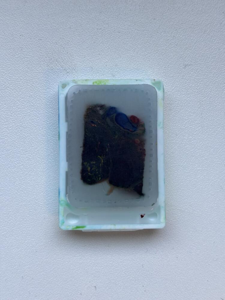
Paraffine kent toepassingen in verschillende domeinen. Zo wordt het gebruikt als omhulsel voor kaas en voor de vervaardiging van kaarsen. In de medische wereld wordt dit gebruikt voor weefselinbedding en is het kenmerkend wit en geurloos. Het is een bijzonder geschikt bewaarmiddel wegens zijn affiniteit met cel- en weefselonderdelen. Een stukje orgaan wordt gefixeerd, ontwaterd en in vloeibare paraffine gegoten, zodat het jarenlang zijn integriteit behoudt.
Een laborant snijdt er plakjes van enkele micrometers, zo’n miljoenste van een meter, af. Ter vergelijking is een micrometer zo’n 50 tot 200 keer fijner dan de dikte van een haar. Deze gesneden plakjes worden dan typisch “gekleurd” met chemicaliën en onder de microscoop bekeken. Deze methode, gekend als histologie, speelt een onmisbare rol bij ziekteleer.
Echter is histologie arbeidsintensief en destructief doordat het snijden de structuur van het weefsel vernietigt. Bovendien levert dit vaak enkel 2D “platte land” informatie op. In het geval dat 3D-informatie vereist is, moet deze methode herhaald en gereconstrueerd worden uit de losse plakjes. Zo is klassieke histologie vergelijkbaar met een brood in dunne plakjes snijden of een roos laag per laag ontvouwen om deze vervolgens te bestuderen.
Fasecontrastbeeldvorming
Wellicht heb je ooit van röntgenstraling gehoord of herinner je die ene keer toen jouw kennis of zielsverwant een pols brak en een “scan” moest nemen. Zo’n röntgenstraal zit ook ingebakken in een RX-toestel of CT-toestel. Röntgenstralen kunnen het blokje paraffine penetreren en leveren beelden op met een enorm goed detail. Echter is het probleem dat paraffine zich tegenover straling bijna hetzelfde gedraagt als het ingebedde weefsel.
Eén oplossing hiervoor is fasecontrast-CT. Röntgenstralen, net als alle andere elektromagnetische golven (in de volksmond bekend als licht), worden niet alleen tegengehouden, maar kunnen ook afgebogen worden wanneer ze de overgang maken van het ene medium naar het andere. Denk hierbij aan water in een doorzichtig glas. Water en glas zijn beide doorzichtig, maar toch zie je het water in het glas door breking. Hetzelfde effect treedt op bij glinsterende lucht boven een wegdek op een hete zomerdag. Dit effect nemen we waar via fasecontrastbeeldvorming van in paraffine ingebedde weefsels. Hierbij maken we gebruik van computeralgoritmes om dit effect om te zetten in een contrastrijk beeld.
Om fasecontrastmetingen te doen, is een heldere röntgenbron vereist. Tot voor kort werd deze techniek beperkt gebruikt bij synchrotrons. Dit zijn deeltjesversnellers met een omtrek van de orde van honderden meters. Toegang tot dergelijke bronnen is beperkt en de stralingsbundel is vaak te klein.
Röntgenbron met vloeibaar doelmateriaal
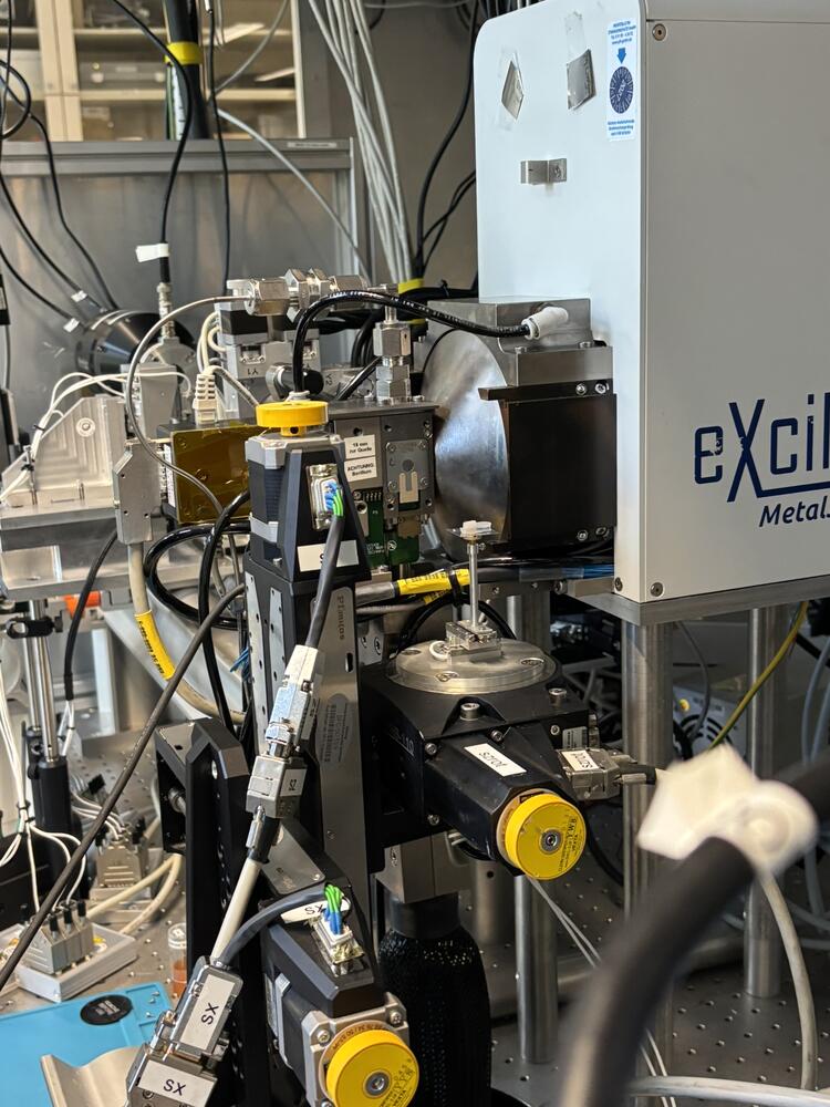
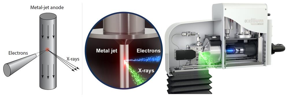
Tijdens mijn masterproef maakte ik aan het Institut für Röntgenphysik in Göttingen gebruik van een alternatieve bron. Conventioneel worden röntgenstralen, zoals Wilhelm Röntgen ze ontdekte eind 19e eeuw, gegenereerd door elektronen op een doelmateriaal (anode) te laten vallen. Typisch stijgt de helderheid van de bron met het aantal invallende elektronen, maar dit gaat gepaard met verhitting, waardoor koeling noodzakelijk is om smelten te voorkomen.
In een liquid-metal jet röntgenbron is het doelmateriaal een dunne straal vloeibaar metaal bestaande uit een legering van gallium, indium en tin. Deze legering, genaamd Galinstan, is vloeibaar bij kamertemperatuur en wordt onder hoge druk rondgepompt, waardoor ze ook continu “ververst” wordt. Op deze manier is de bron in staat om een pak helderdere straling te leveren dan een klassieke röntgenbuis.
Kleur speelt naast felheid ook een belangrijke rol
Tijdens mijn thesis stelde ik het systeem af, gaande van afstanden en positionering tot rekenmethodes. Het resultaat is een recept om 3D-beeldvorming te doen op in paraffine ingebedde weefsels. Details van zo’n 15 micrometer, ongeveer zo groot als een wit bloedlichaampje, konden op betrouwbare wijze waargenomen worden. Ik bestuurde weefsels uit de luchtwegen, hersenen, bloedvaten en placenta’s. In de hippocampus van een patiënt met epilepsie konden verschillende regio’s in het weefsel worden onderscheiden. De placenta werd vóór het inbedden met contrastvloeistof gekleurd. Op de scans waren bloedvaten zichtbaar, maar ook een fijn netwerk waarlangs de moeder en het kind stoffen met elkaar uitwisselden.
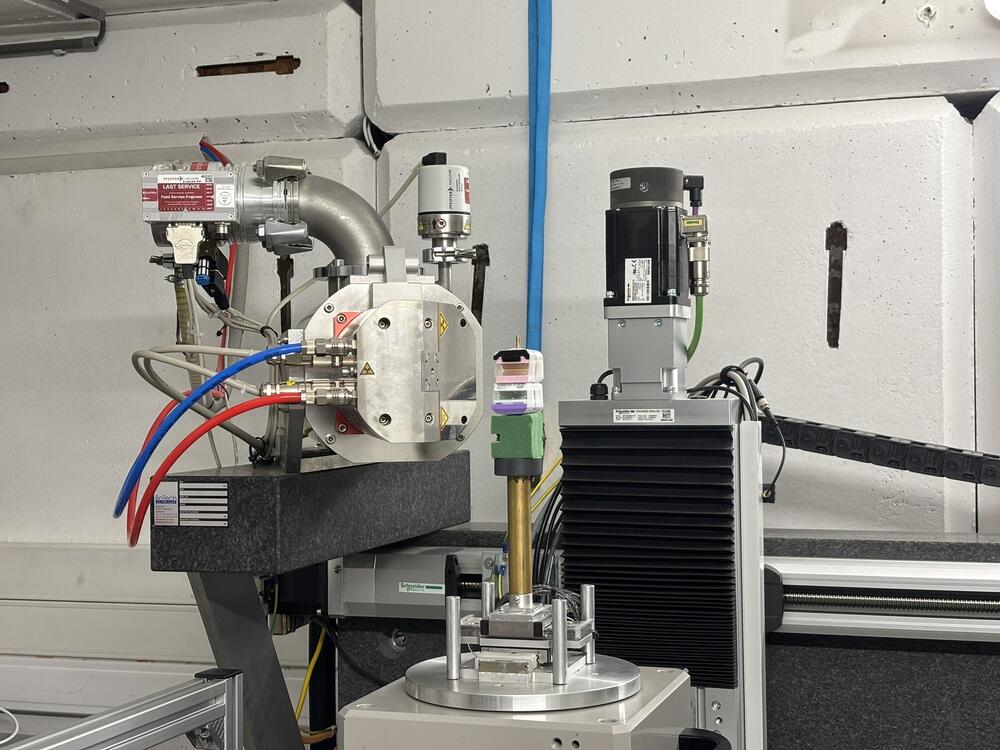
Ter vergelijking werd ook een scan uitgevoerd met andere CT-toestellen, aan UGCT in Gent en in Göttingen. Deze werken met meer energierijke straling die vlotter door het blokje heen gaat. Een belangrijke les gaat over kleur. Röntgenstraling, net als een regenboog, bestaat uit verschillende kleuren (energieën), uitgedrukt in kilo-elektronvolt (keV). De felste kleur (gallium op 9 keV) van de liquid-metal jet bron werd grotendeels geabsorbeerd door de paraffine, terwijl een relatief groot deel van het werk door indium (24 keV) werd gedaan. Een vierde toestel, met een vast molybdeendoelmateriaal en een vermogen van amper een fractie van de liquid-metal jet (4 watt in plaats van 57 watt), haalde in bepaalde gebieden een hoger contrast.
Molybdeen straalt zijn dominante kleur bij 17,5 keV precies in een zone waar het hoog genoeg is om doorheen paraffine te gaan, maar ook niet te hoog om het contrast te doen zakken. Een ideale bron voor blokjes van die grootte bevindt zich vooral tussen ongeveer 15 en 25 keV. Een liquid-metal jet bron met een hoog indiumgehalte kan een interessante kandidaat zijn. Samen met detectors (camera's) die hierop beter afgestemd zijn, kan de scanduur aanzienlijk worden verlaagd.
Eerst digitaal binnenkijken, dan snijden
Met dergelijke studies en opkomende technieken komt “virtuele” histologie binnen het bereik van gewone labo’s en binnenkort hopelijk ook in ziekenhuizen. Voordelen van deze techniek zijn dat het blokje intact blijft, beeldvorming snel kan gebeuren en het in 3D bestudeerd kan worden. De in paraffine ingebedde blokjes kunnen indien nodig altijd na het scannen gesneden, gekleurd en klassiek waargenomen worden. Bovendien kan men zich met fasecontrastbeeldvorming specifiek richten op plaatsen waar het interessant lijkt om te snijden en op hoe dit moet gebeuren.
Bibliografie
Bibliografie
@phdthesis{booneNewImagingModalities2013,
type = {dissertation},
title = {New imaging modalities in high resolution {X}-ray tomography},
copyright = {No license (in copyright)},
url = {http://hdl.handle.net/1854/LU-4157322},
abstract = {For more than 100 years, X-rays are used in medical diagnostics, materials science and many other research domains for the possibility they offer to look through objects. Since the 1970s, Computed Tomography or CT allows for acquiring a full 3D representation of an object or a patient. In the 1980s, technological advances led to the development of high resolution X-ray CT or µCT.
Using this technique, objects can be investigated with a spatial resolution ranging from several tens of micrometers to less than one micrometer. Despite the strong evolution in µCT, many challenges remain. Apart from several generic developments contributing to the improvement of the technique, two new imaging modalities are being investigated in this dissertation. The first modality accounts for the refraction of X-rays which contributes, along with X-ray attenuation, to the observed image. This modality is called X-ray phase contrast imaging. The second modality accounts for the energy-dependent attenuation of X-rays and allows to obtain more chemical information on the object under investigation. This modality is called spectral absorption tomography.
In X-ray phase contrast imaging, the observed image contains contributions from both X-ray refraction and X-ray attenuation. Because there is no unique correlation between both effects, it is impossible to decouple both contributions from one single image, which gives rise to imaging artifacts in a classical CT reconstruction. Special reconstruction algorithms are thus required to obtain an approximated solution and to reduce imaging artifacts. In this dissertation, 4 such algorithms are elaborated and characterized based on both simulated and experimental data.
In spectral absorption tomography, different images are recorded at different spectral characteristics. One method to achieve this is the usage of energy-dispersive detector systems. In this work, two such systems are discussed, using a different methodology to obtain the spectral information. The advantages and limitations of both systems are discussed and tested in the scope of acquiring three-dimensional chemical information non-destructively.},
language = {eng},
urldate = {2024-12-21},
school = {Ghent University},
author = {Boone, Matthieu},
year = {2013},
note = {ISBN: 9789461971494}
}
@book{saldittBiomedicalImagingPrinciples2017,
title = {Biomedical {Imaging}: {Principles} of {Radiography}, {Tomography} and {Medical} {Physics}},
isbn = {978-3-11-042669-4},
shorttitle = {Biomedical {Imaging}},
abstract = {Covering both physical as well as mathematical and algorithmic foundations, this graduate textbook provides the reader with an introduction into modern biomedical imaging and image processing and reconstruction. These techniques are not only based on advanced instrumentation for image acquisition, but equally on new developments in image processing and reconstruction to extract relevant information from recorded data. To this end, the present book offers a quantitative treatise of radiography, computed tomography, and medical physics. ContentsIntroductionDigital image processingEssentials of medical x-ray physicsTomographyRadiobiology, radiotherapy, and radiation protectionPhase contrast radiographyObject reconstruction under nonideal conditions},
language = {en},
publisher = {Walter de Gruyter GmbH \& Co KG},
author = {Salditt, Tim and Aspelmeier, Timo and Aeffner, Sebastian},
month = oct,
year = {2017},
note = {Google-Books-ID: nMA7DwAA
QBAJ},
keywords = {Computers / Data Science / Data Analytics, Computers / Data Science / General, Computers / Database Administration \& Management, Computers / Optical Data Processing, Mathematics / Applied, Mathematics / Numerical Analysis, Medical / Allied Health Services / Imaging Technologies, Medical / Radiology, Radiotherapy \& Nuclear Medicine, Science / Physics / General, Science / Physics / Optics \& Light}
}
@misc{mertensKwantumveldenTheorie2023,
title = {Kwantumvelden theorie},
shorttitle = {{QFT}},
language = {Dutch},
publisher = {UGent},
author = {Mertens, Thomas and Verschelde, Henri},
year = {2023}
}
@misc{enwikiOpticaltransferfunction,
author = {{Wikipedia contributors}},
title = {Optical transfer function --- {Wikipedia}{,} The Free Encyclopedia},
year = {2025},
url = {https://en.wikipedia.org/w/index.php?title=Optical_transfer_function&ol…},
note = {[Online; accessed 19-September-2025]}
}
@misc{joosAtomicMolecularPhysics2024,
title = {Atomic and {Molecular} {Physics}},
language = {English},
publisher = {UGent},
author = {Joos, Jonas and Vrielinck, Henk},
year = {2024}
}
@misc{JuliaIrp,
title = {julia [irp]},
url = {https://irp.wiki.gwdg.de/doku.php?id=julia},
language = {English},
urldate = {2024-12-22},
journal = {Wiki IRP Göttingen}
}
@misc{thomsonModernParticlePhysics2013,
title = {Modern {Particle} {Physics}},
url = {https://www.cambridge.org/highereducation/books/modern-particle-physics…},
abstract = {Unique in its coverage of all aspects of modern particle physics, this textbook provides a clear connection between the theory and recent experimental results, including the discovery of the Higgs boson at CERN. It provides a comprehensive and self-contained description of the Standard Model of particle physics suitable for upper-level undergraduate students and graduate students studying experimental particle physics. Physical theory is introduced in a straightforward manner with full mathematical derivations throughout. Fully-worked examples enable students to link the mathematical theory to results from modern particle physics experiments. End-of-chapter exercises, graded by difficulty, provide students with a deeper understanding of the subject. Online resources available at www.cambridge.org/MPP feature password-protected fully-worked solutions to problems for instructors, numerical solutions and hints to the problems for students and PowerPoint slides and JPEGs of figures from the book.},
language = {en-BE},
urldate = {2024-12-22},
journal = {Higher Education from Cambridge University Press},
author = {Thomson, Mark},
month = sep,
year = {2013},
doi = {10.1017/CBO9781139525367},
note = {ISBN: 9781139525367
Publisher: Cambridge University Press}
}
@book{barrettFoundationsImageScience2013,
title = {Foundations of {Image} {Science}},
isbn = {978-1-118-77277-5},
abstract = {Winner of the 2006 Joseph W. Goodman Book Writing Award! A comprehensive treatment of the principles, mathematics, and statistics of image science In today's visually oriented society, images play an important role in conveying messages. From seismic imaging to satellite images to medical images, our modern society would be lost without images to enhance our understanding of our health, our culture, and our world. Foundations of Image Science presents a comprehensive treatment of the principles, mathematics, and statistics needed to understand and evaluate imaging systems. The book is the first to provide a thorough treatment of the continuous-to-discrete, or CD, model of digital imaging. Foundations of Image Science emphasizes the need for meaningful, objective assessment of image quality and presents the necessary tools for this purpose. Approaching the subject within a well-defined theoretical and physical context, this landmark text presents the mathematical underpinnings of image science at a level that is accessible to graduate students and practitioners working with imaging systems, as well as well-motivated undergraduate students. Destined to become a standard text in the field, Foundations of Image Science covers: Mathematical Foundations: Examines the essential mathematical foundations of image science Image Formation–Models and Mechanisms: Presents a comprehensive and unified treatment of the mathematical and statistical principles of imaging, with an emphasis on digital imaging systems and the use of SVD methods Image Quality: Provides a systematic exposition of the methodology for objective or task-based assessment of image quality Applications: Presents detailed case studies of specific direct and indirect imaging systems and provides examples of how to apply the various mathematical tools covered in the book Appendices: Covers the prerequisite material necessary for understanding the material in the main text, including matrix algebra, complex variables, and the basics of probability theory},
language = {en},
publisher = {John Wiley \& Sons},
author = {Barrett, Harrison H. and Myers, Kyle J.},
month = jun,
year = {2013},
note = {Google-Books-ID: XCypDoCKtQMC},
keywords = {Science / Physics / Atomic \& Molecular, Technology \& Engineering / Electrical, Technology \& Engineering / Electronics / General}
}
@article{kuhls-gilcristAccurateMTFMeasurement2010,
title = {Accurate {{MTF}} Measurement in Digital Radiography Using Noise Response},
author = {Kuhls-Gilcrist, Andrew and Jain, Amit and Bednarek, Daniel R. and Hoffmann, Kenneth R. and Rudin, Stephen},
date = {2010-02},
journaltitle = {Medical Physics},
shortjournal = {Med Phys},
volume = {37},
number = {2},
eprint = {20229882},
eprinttype = {pmid},
pages = {724--735},
issn = {0094-2405},
doi = {10.1118/1.3284376},
url = {https://www.ncbi.nlm.nih.gov/pmc/articles/PMC2821422/},
urldate = {2024-12-20},
abstract = {Purpose: The authors describe a new technique to determine the system presampled modulation transfer function (MTF) in digital radiography using only the detector noise response., Methods: A cascaded-linear systems analysis was used to develop an exact relationship between the two-dimensional noise power spectrum (NPS) and the presampled MTF for a generalized detector system. This relationship was then utilized to determine the two-dimensional presampled MTF. For simplicity, aliasing of the correlated noise component of the NPS was assumed to be negligible. Accuracy of this method was investigated using simulated images from a simple detector model in which the “true” MTF was known exactly. Measurements were also performed on three detector technologies (an x-ray image intensifier, an indirect flat panel detector, and a solid state x-ray image intensifier), and the results were compared using the standard edge-response method. Flat-field and edge images were acquired and analyzed according to guidelines set forth by the International Electrotechnical Commission, using the RQA 5 spectrum., Results: The presampled MTF determined using the noise-response method for the simulated detector system was in close agreement with the true MTF with an averaged percent difference of 0.3\% and a maximum difference of 1.1\% observed at the Nyquist frequency (fN). The edge-response method of the simulated detector system also showed very good agreement at lower spatial frequencies (less than 0.5 fN) with an averaged percent difference of 1.6\% but showed significant discrepancies at higher spatial frequencies (greater than 0.5 fN) with an averaged percent difference of 17\%. Discrepancies were in part a result of noise in the edge image and phasing errors. For all three detector systems, the MTFs obtained using the two methods were found to be in good agreement at spatial frequencies less than 0.5 fN with an averaged percent difference of 3.4\%. Above 0.5 fN, differences increased to an average of 20\%. Deviations of the experimental results largely followed the trend seen in the simulation results, suggesting that differences between the two methods could be explained as resulting from the inherent inaccuracies of the edge-response measurement technique used in this study. Aliasing of the correlated noise component was shown to have a minimal effect on the measured MTF for the three detectors studied. Systems with significant aliasing of the correlated noise component (e.g., a-Se based detectors) would likely require a more sophisticated fitting scheme to provide accurate results., Conclusions: Results indicate that the noise-response method, a simple technique, can be used to accurately measure the MTF of digital x-ray detectors, while alleviating the problems and inaccuracies associated with use of precision test objects, such as a slit or an edge.},
pmcid = {PMC2821422}
}
@misc{enwikidynamicflatfieldcorrection,
author = {{Wikipedia contributors}},
title = {Flat-field correction --- {Wikipedia}{,} The Free Encyclopedia},
year = {2024},
url = {https://en.wikipedia.org/w/index.php?title=Flat-field_correction&oldid=…},
note = {[Online; accessed 14-April-2025]}
}
@misc{enwikiBremsstrahlung,
author = {{Wikipedia contributors}},
title = {Bremsstrahlung --- {Wikipedia}{,} The Free Encyclopedia},
year = {2025},
url = {https://en.wikipedia.org/w/index.php?title=Bremsstrahlung&oldid=1284460…},
note = {[Online; accessed 17-April-2025]}
}
@article{por2019nyquist,
title = {Nyquist--Shannon sampling theorem},
author = {Por, Emiel and Van Kooten, Maaike and Sarkovic, Vanja},
journal = {Leiden University},
volume = {1},
number = {1},
pages = {1--2},
year = {2019}
}
@misc{enwikiKramersLaw,
author = {{Wikipedia contributors}},
title = {Kramers' law --- {Wikipedia}{,} The Free Encyclopedia},
year = {2023},
url = {https://en.wikipedia.org/w/index.php?title=Kramers%27_law&oldid=1174532…},
note = {[Online; accessed 24-April-2025]}
}
@phdthesis{pauwelsOptimalUsePolychromaticity2017,
type = {dissertation},
title = {Optimal use of the polychromaticity in {X}-ray microtomography for dual energy methods and contrast agent imaging},
copyright = {No license (in copyright)},
url = {http://hdl.handle.net/1854/LU-8514881},
language = {eng},
urldate = {2025-04-17},
school = {Ghent University},
author = {Pauwels, Elin},
year = {2017},
note = {ISBN: 9789461975027}
}
@misc{enwikiDuaneHuntLaw,
author = {{Wikipedia contributors}},
title = {Duane–Hunt law --- {Wikipedia}{,} The Free Encyclopedia},
year = {2024},
url = {https://en.wikipedia.org/w/index.php?title=Duane%E2%80%93Hunt_law&oldid…},
note = {[Online; accessed 24-April-2025]}
}
@misc{CXRO2025,
title = {{The index of refraction for a compound material}},
author = {{The Center for X-ray Optics}},
year = {2025},
note = {Accessed: 2025-05-01},
url = {https://henke.lbl.gov/optical_constants/}
}
@misc{enwikiParaffin,
author = {{Wikipedia contributors}},
title = {Paraffin wax --- {Wikipedia}{,} The Free Encyclopedia},
year = {2024},
url = {https://en.wikipedia.org/w/index.php?title=Paraffin_wax&oldid=1258851351},
note = {[Online; accessed 1-May-2025]}
}
@misc{enwikivanderWaalsForce,
author = {{Wikipedia contributors}},
title = {Van der Waals force --- {Wikipedia}{,} The Free Encyclopedia},
year = {2025},
url = {https://en.wikipedia.org/w/index.php?title=Van_der_Waals_force&oldid=12…},
note = {[Online; accessed 1-May-2025]}
}
@misc{PubChem_Paraffin,
title = {{Paraffin}},
author = {{National Center for Biotechnology Information}},
year = {2025},
howpublished = {PubChem Compound Database},
note = {Accessed: 2025-05-01},
url = {https://pubchem.ncbi.nlm.nih.gov/compound/Paraffin}
}
@misc{CarlRoth_Paraplast,
title = {{Paraplast - Paraffin for Tissue Embedding}},
author = {{Carl ROTH GmbH}},
year = {2025},
howpublished = {Product Information Sheet},
note = {Accessed: 2025-05-01},
url = {https://www.carlroth.com/de/de/paraplast-paraffine/paraplast/p/x880.1}
}
@misc{enwikiMaxwellequations,
author = {{Wikipedia contributors}},
title = {Maxwell's equations --- {Wikipedia}{,} The Free Encyclopedia},
year = {2025},
url = {https://en.wikipedia.org/w/index.php?title=Maxwell%27s_equations&oldid=…},
note = {[Online; accessed 9-June-2025]}
}
@book{griffiths2023introduction,
title = {Introduction to electrodynamics},
author = {Griffiths, David J},
year = {2023},
publisher = {Cambridge University Press}
}
@book{katsenelenbaumHighfrequencyElectrodynamics2006,
title = {High-Frequency Electrodynamics},
author = {Katsenelenbaum, Boris Zakharovich},
date = {2006},
publisher = {Wiley-VCH},
location = {Weinheim},
isbn = {978-3-527-40529-9},
langid = {english},
url = {https://application.wiley-vch.de/books/sample/3527405291_c01.pdf}
}
@book{paganin2006coherent,
title = {Coherent X-ray optics},
author = {Paganin, David},
number = {6},
year = {2006},
publisher = {Oxford University Press},
doi = {10.1093/acprof:oso/9780198567288.001.0001},
abstract = {This book offers a grounding in the field of coherent X-ray optics, which in the closing years of the 20th century experienced something of a renaissance with the availability of third-generation synchrotron sources. It begins with a treatment of the fundamentals of X-ray diffraction for both coherent and partially coherent radiation, together with the interactions of X-rays with matter. X-ray sources, optical elements, and detectors are then discussed, with an emphasis on their role in coherent X-ray optics. Various aspects of coherent X-ray imaging are then considered, including holography, interferometry, self imaging, phase contrast, and phase retrieval. The foundations of the new field of singular X-ray optics are examined, focusing on the topic of X-ray phase vortices. Most topics in the book are developed from first principles using a chain of logic which ultimately derives from the Maxwell equations, with numerous references to the contemporary and historical research literature.},
isbn = {978-0-19-856728-8}
}
@misc{enwikiDensityFunctionalTheory,
author = {{Wikipedia contributors}},
title = {Density functional theory --- {Wikipedia}{,} The Free Encyclopedia},
year = {2025},
url = {https://en.wikipedia.org/w/index.php?title=Density_functional_theory&ol…},
note = {[Online; accessed 9-June-2025]}
}
@misc{enwikiEnergyPhoton,
author = {{Wikipedia contributors}},
title = {Photon energy --- {Wikipedia}{,} The Free Encyclopedia},
year = {2024},
url = {https://en.wikipedia.org/w/index.php?title=Photon_energy&oldid=12585778…},
note = {[Online; accessed 9-June-2025]}
}
@online{X123CompleteXRay,
title = {X-123 {{Complete X-Ray Spectrometer}} with {{Si-PIN Detector}} – {{Amptek}} – {{X-Ray Detectors}} and {{Electronics}}},
url = {https://www.amptek.com/internal-products/x-123-complete-x-ray-spectrome…},
urldate = {2025-06-09},
abstract = {X-123 Complete X-Ray Spectrometer with Si-PIN Detector},
langid = {english}
}
@misc{enwikiLagrangianFieldTheory,
author = {{Wikipedia contributors}},
title = {Lagrangian (field theory) --- {Wikipedia}{,} The Free Encyclopedia},
year = {2025},
url = {https://en.wikipedia.org/w/index.php?title=Lagrangian_(field_theory)&ol…},
note = {[Online; accessed 9-June-2025]}
}
@misc{enwikiTEM,
author = {{Wikipedia contributors}},
title = {Transmission electron microscopy --- {Wikipedia}{,} The Free Encyclopedia},
year = {2025},
url = {https://en.wikipedia.org/w/index.php?title=Transmission_electron_micros…},
note = {[Online; accessed 10-June-2025]}
}
@misc{tomasi2017image,
title = {Image correlation, convolution and filtering},
author = {Tomasi, Carlo}
}
@misc{enwikiPhaseCorrelation,
author = {{Wikipedia contributors}},
title = {Phase correlation --- {Wikipedia}{,} The Free Encyclopedia},
year = {2024},
url = {https://en.wikipedia.org/w/index.php?title=Phase_correlation&oldid=1265…},
note = {[Online; accessed 11-June-2025]}
}
@misc{enwikiIntegralTransform,
author = {{Wikipedia contributors}},
title = {Integral transform --- {Wikipedia}{,} The Free Encyclopedia},
year = {2024},
url = {https://en.wikipedia.org/w/index.php?title=Integral_transform&oldid=125…},
note = {[Online; accessed 14-June-2025]}
}
@article{de2009bronnikov,
title = {Bronnikov-aided correction for x-ray computed tomography},
author = {De Witte, Yoni and Boone, Matthieu and Vlassenbroeck, Jelle and Dierick, Manuel and Van Hoorebeke, Luc},
journal = {Journal of the Optical Society of America A},
volume = {26},
number = {4},
pages = {890--894},
year = {2009},
publisher = {Optical Society of America}
}
@misc{lucht2025hotopytoolboxxrayholotomography,
title = {HoToPy: A toolbox for X-ray holo-tomography in Python},
author = {Jens Lucht and Paul Meyer and Leon Merten Lohse and Tim Salditt},
year = {2025},
eprint = {2506.11567},
archiveprefix = {arXiv},
primaryclass = {physics.optics},
url = {https://arxiv.org/abs/2506.11567}
}
@inbook{inbookoctopus,
author = {Vlassenbroeck, Jelle and Masschaele, Bert and Cnudde, Veerle and Dierick, Manuel and Pieters, Koen and Hoorebeke, Luc and Jacobs, Patric},
year = {2010},
month = {01},
pages = {167-173},
title = {Octopus 8: A High Performance Tomographic Reconstruction Package for X‐ray Tube and Synchrotron micro‐CT},
isbn = {9781905209606},
doi = {10.1002/9780470612187.ch13}
}
@book{turbell2001cone,
title = {Cone-beam reconstruction using filtered backprojection},
author = {Turbell, Henrik},
year = {2001},
publisher = {Linkopings Universitet (Sweden)}
}
@article{Feldkamp:84,
author = {L. A. Feldkamp and L. C. Davis and J. W. Kress},
journal = {J. Opt. Soc. Am. A},
keywords = {Destructive interference; Detector arrays; Fourier transforms; Systems design; Three dimensional reconstruction; X-ray imaging},
number = {6},
pages = {612--619},
publisher = {Optica Publishing Group},
title = {Practical cone-beam algorithm},
volume = {1},
month = {6},
year = {1984},
url = {https://opg.optica.org/josaa/abstract.cfm?URI=josaa-1-6-612},
doi = {10.1364/JOSAA.1.000612},
abstract = {A convolution-backprojection formula is deduced for direct reconstruction of a three-dimensional density function from a set of two-dimensional projections. The formula is approximate but has useful properties, including errors that are relatively small in many practical instances and a form that leads to convenient computation. It reduces to the standard fan-beam formula in the plane that is perpendicular to the axis of rotation and contains the point source. The algorithm is applied to a mathematical phantom as an example of its performance.}
}
@article{virtualhistology2024,
author = {Tajbakhsh, Kiarash and Neels, Antonia and Fadeeva, Elena and Larsson, Jakob and Stanowska, Olga and Perren, Aurel and Zboray, Robert},
year = {2024},
month = {03},
pages = {},
title = {A comprehensive study of micro-CT for 3D virtual histology of FFPE tissue blocks},
doi = {10.36227/techrxiv.171177300.04278071/v1}
}
@misc{enwikiLevenbergMarquardtAlgorithm,
author = {{Wikipedia contributors}},
title = {Levenberg–Marquardt algorithm --- {Wikipedia}{,} The Free Encyclopedia},
year = {2024},
url = {https://en.wikipedia.org/w/index.php?title=Levenberg%E2%80%93Marquardt_…},
note = {[Online; accessed 23-June-2025]}
}
@misc{dewikiRontgen,
author = {Wikipedia},
title = {Wilhelm Conrad Röntgen --- Wikipedia{,} the free encyclopedia},
year = {2025},
url = { https://de.wikipedia.org/w/index.php?title=Wilhelm_Conrad_R%C3%B6ntgen&… },
note = {[Online; as of August 11, 2025]}
}
@article{paganinphaseAmplitudeExtraction2002,
title = {Simultaneous Phase and Amplitude Extraction from a Single Defocused Image of a Homogeneous Object},
author = {Paganin, {David Maurice} and Mayo, {Sheridan C} and Gureyev, {Timur E} and Miller, {Peter Robert} and Wilkins, {S W}},
year = {2002},
language = {English},
volume = {206},
pages = {33 -- 40},
journal = {Journal of Microscopy},
issn = {0022-2720},
publisher = {Wiley-Blackwell},
number = {1}
}
@article{paganin2019tutorials,
title = {Tutorials on x-ray phase contrast imaging: Some fundamentals and some conjectures on future developments},
author = {Paganin, David M and Pelliccia, Daniele},
journal = {arXiv preprint arXiv:1902.00364},
year = {2019}
}
@article{Masschaele_2013,
doi = {10.1088/1742-6596/463/1/012012},
url = {https://dx.doi.org/10.1088/1742-6596/463/1/012012},
year = {2013},
month = {10},
publisher = {},
volume = {463},
number = {1},
pages = {012012},
author = {Masschaele, Bert and Dierick, Manuel and Loo, Denis Van and Boone, Matthieu N and Brabant, Loes and Pauwels, Elin and Cnudde, Veerle and Hoorebeke, Luc Van},
title = {HECTOR: A 240kV micro-CT setup optimized for research},
journal = {Journal of Physics: Conference Series},
abstract = {X-ray micro-CT has become a very powerful and common tool for non-destructive three-dimensional (3D) visualization and analysis of objects. Many systems are commercially available, but they are typically limited in terms of operational freedom both from a mechanical point of view as well as for acquisition routines. HECTOR is the latest system developed by the Ghent University Centre for X-ray Tomography (http://www.ugct.ugent.be) in collaboration with X-Ray Engineering (XRE bvba, Ghent, Belgium). It consists of a mechanical setup with nine motorized axes and a modular acquisition software package and combines a microfocus directional target X-ray source up to 240 kV with a large flat-panel detector. Provisions are made to install a line-detector for a maximal operational range. The system can accommodate samples up to 80 kg, 1 m long and 80 cm in diameter while it is also suited for high resolution (down to 4 μm) tomography. The bi-directional detector tiling is suited for large samples while the variable source-detector distance optimizes the signal to noise ratio (SNR) for every type of sample, even with peripheral equipment such as compression stages or climate chambers. The large vertical travel of 1 m can be used for helical scanning and a vertical detector rotation axis allows laminography experiments.
The setup is installed in a large concrete bunker to allow accommodation of peripheral equipment such as pumps, chillers, etc., which can be integrated in the modular acquisition software to obtain a maximal correlation between the environmental control and the CT data taken. The acquisition software does not only allow good coupling with the peripheral equipment but its scripting feature is also particularly interesting for testing new and exotic acquisition routines.}
}
@misc{xrayworx2025,
author = {{X-Ray WorX GmbH}},
title = {High-resolution microfocus X-ray tubes},
year = {2025},
note = {Accessed: 2025-06-26},
url = {https://www.x-ray-worx.com/}
}
@article{harris2020array,
title = {Array programming with {NumPy}},
author = {Charles R. Harris and K. Jarrod Millman and St{\'{e}}fan J.
van der Walt and Ralf Gommers and Pauli Virtanen and David
Cournapeau and Eric Wieser and Julian Taylor and Sebastian
Berg and Nathaniel J. Smith and Robert Kern and Matti Picus
and Stephan Hoyer and Marten H. van Kerkwijk and Matthew
Brett and Allan Haldane and Jaime Fern{\'{a}}ndez del
R{\'{i}}o and Mark Wiebe and Pearu Peterson and Pierre
G{\'{e}}rard-Marchant and Kevin Sheppard and Tyler Reddy and
Warren Weckesser and Hameer Abbasi and Christoph Gohlke and
Travis E. Oliphant},
year = {2020},
month = sep,
journal = {Nature},
volume = {585},
number = {7825},
pages = {357--362},
doi = {10.1038/s41586-020-2649-2},
publisher = {Springer Science and Business Media {LLC}},
url = {https://doi.org/10.1038/s41586-020-2649-2}
}
@misc{enwikiXraytube,
author = {{Wikipedia contributors}},
title = {X-ray tube --- {Wikipedia}{,} The Free Encyclopedia},
year = {2025},
url = {https://en.wikipedia.org/w/index.php?title=X-ray_tube&oldid=1297675252},
note = {[Online; accessed 28-June-2025]}
}
@misc{excillumhistoryxraytubes,
author = {Bernard, David},
title = {History of X-rays 125 years in the making (pt 2)},
url = {https://www.excillum.com/history-of-x-rays-x-ray-tubes/},
institution = {Excillum AB},
note = {Accessed: 2025-06-28},
year = {2020},
month = {10},
day = {8}
}
@misc{rigaku2025beamhardening,
title = {What Is Beam Hardening in CT?},
author = {Takase Aya},
url = {https://rigaku.com/products/imaging-ndt/x-ray-ct/learning/blog/what-is-…},
year = {2025},
month = {9},
day = {30},
note = {Rigaku Corporation. Accessed: 2025-06-28}
}
@article{Croton:19,
author = {Linda C. P. Croton and Gary Ruben and Kaye S. Morgan and David M. Paganin and Marcus J. Kitchen},
journal = {Opt. Express},
keywords = {Attenuation coefficient; Image quality; Phase contrast; Phase imaging; Point spread function; X-ray imaging},
number = {10},
pages = {14231--14245},
publisher = {Optica Publishing Group},
title = {Ring artifact suppression in X-ray computed tomography using a simple, pixel-wise response correction},
volume = {27},
month = {5},
year = {2019},
url = {https://opg.optica.org/oe/abstract.cfm?URI=oe-27-10-14231},
doi = {10.1364/OE.27.014231},
abstract = {We present a pixel-specific, measurement-driven correction that effectively reduces errors in detector response that give rise to the ring artifacts commonly seen in X-ray computed tomography (CT) scans. This correction is easy to implement, suppresses CT artifacts significantly, and is effective enough for use with both absorption and phase contrast imaging. It can be used as a standalone correction or in conjunction with existing ring artifact removal algorithms to further improve image quality. We validate this method using two X-ray CT data sets acquired using monochromatic sources, showing post-correction signal-to-noise increases of up to 55\&\#x00025;, and we define an image quality metric to use specifically for the assessment of ring artifact suppression.}
}
@misc{shi2024ringartifactsremovalbased,
title = {Ring Artifacts Removal Based on Implicit Neural Representation of Sinogram Data},
author = {Ligen Shi and Xu Jiang and YunZe Liu and Chang Liu and Ping Yang and Shifeng Guo and Xing Zhao},
year = {2024},
eprint = {2409.15731},
archiveprefix = {arXiv},
primaryclass = {eess.IV},
url = {https://arxiv.org/abs/2409.15731}
}
@article{Yang:20,
author = {Yafei Yang and Dinghua Zhang and Fuqiang Yang and Mingxuan Teng and You Du and Kuidong Huang},
journal = {Opt. Express},
keywords = {Computed tomography; Deep learning; Fourier transforms; Numerical simulation; Photon counting; Spatial resolution},
number = {21},
pages = {30362--30378},
publisher = {Optica Publishing Group},
title = {Post-processing method for the removal of mixed ring artifacts in CT images},
volume = {28},
month = {10},
year = {2020},
url = {https://opg.optica.org/oe/abstract.cfm?URI=oe-28-21-30362},
doi = {10.1364/OE.401088},
abstract = {Ring artifacts seriously deteriorate the quality of CT images. Intensity-dependence of detector responses will result in intensity-dependent ring artifacts and time-dependence of CT hardware systems will result in time-dependent ring artifacts. However, only the intensity-dependent ring artifacts are taken into consideration in most post-processing methods. Therefore, the purpose of this study is to propose a general post-processing method, which has a significant removal effect on the intensity-dependent ring artifacts and the time-dependent ring artifacts. First in the proposed method, transform raw CT images into polar coordinate images, and the ring artifacts will manifest as stripe artifacts. Secondly, obtain structure images by smoothing the polar coordinate images and acquire texture images containing some details and stripe artifacts by subtracting the structure images from the polar coordinate images. Third, extract the stripe artifacts from the texture images using mean extraction and texture classification, and obtain the extracted ring artifacts by transforming the extracted stripe artifacts from polar coordinates into Cartesian coordinates. Finally, obtain corrected CT images by subtracting the extracted ring artifacts from the raw CT images, and iterate the corrected CT images in above steps until the ring artifacts extracted in the last iteration are weak enough. Simulation and real data show that the proposed method can remove the intensity-dependent ring artifacts and the time-dependent ring artifacts effectively while preserving image details and spatial resolution. In particular, real data prove that the method is suitable for new CT systems such as the photon counting CT.}
}
@article{chenPartialditheringStrategyEdgeillumination2020,
title = {A Partial-Dithering Strategy for Edge-Illumination x-Ray Phase-Contrast Tomography Enabled by a Joint Reconstruction Method},
author = {Chen, Yujia and Hagen, Charlotte K. and Olivo, Alessandro and Anastasio, Mark A.},
year = {2020},
month = may,
journal = {Physics in Medicine and Biology},
volume = {65},
number = {10},
pages = {105007},
issn = {1361-6560},
doi = {10.1088/1361-6560/ab66e2},
abstract = {Edge-illumination x-ray phase-contrast tomography (EIXPCT) is a promising imaging technology where partially opaque masks are utilized with laboratory-based x-ray sources to estimate the distribution of the complex-valued refractive index. EIXPCT resolution is mainly determined by the period of a sample mask, but can be significantly improved by a dithering technique. Here, dithering means that multiple images per tomographic view angle are acquired as the object is moved over sub-pixel distances. Drawbacks of dithering include increased data-acquisition times and radiation doses. Motivated by the flexibility in data-acquisition designs enabled by a recently developed joint reconstruction method, a novel partial-dithering strategy for EIXPCT data-acquisition is proposed. In this strategy, dithering is implemented at only a subset of the tomographic view angles. The strategy can result in spatial resolution comparable to that of the conventional full-dithering strategy, where dithering is performed at every view angle, but the acquisition time is substantially decreased. Here, the effect of dithering parameters on image resolution is explored.},
langid = {english},
pmid = {31896094},
keywords = {Image Processing Computer-Assisted,Lighting,Phantoms Imaging,Refractometry,Tomography X-Ray Computed}
}
@misc{wellman2023flatpanel,
author = {Chan Jeff},
institution = {Wellman X-Ray Solution Co., Ltd.},
title = {Everything You Need to Know about Flat Panel Detector},
url = {https://wellmanxray.com/blog/everything-you-need-to-know-about-flat-pan…},
langid = {english},
year = {2023},
month = {5},
day = {31},
note = {Accessed: 2025-06-28}
}
@misc{enwikiX_ray_detector,
author = {{Wikipedia contributors}},
title = {X-ray detector --- {Wikipedia}{,} The Free Encyclopedia},
year = {2025},
url = {https://en.wikipedia.org/w/index.php?title=X-ray_detector&oldid=1268165…},
note = {[Online; accessed 2-July-2025]}
}
@article{verbekeSelfFourierShell2024,
title = {Self {{Fourier}} Shell Correlation: Properties and Application to Cryo-{{ET}}},
author = {Verbeke, Eric J. and Gilles, Marc Aur{\`e}le and Bendory, Tamir and Singer, Amit},
year = {2024},
month = jan,
journal = {Communications Biology},
volume = {7},
number = {1},
pages = {101},
issn = {2399-3642},
doi = {10.1038/s42003-023-05724-y},
abstract = {The Fourier shell correlation (FSC) is a measure of the similarity between two signals computed over corresponding shells in the frequency domain and has broad applications in microscopy. In structural biology, the FSC is ubiquitous in methods for validation, resolution determination, and signal enhancement. Computing the FSC usually requires two independent measurements of the same underlying signal, which can be limiting for some applications. Here, we analyze and extend on an approach to estimate the FSC from a single measurement. In particular, we derive the necessary conditions required to estimate the FSC from downsampled versions of a single noisy measurement. These conditions reveal additional corrections which we implement to increase the applicability of the method. We then illustrate two applications of our approach, first as an estimate of the global resolution from a single 3-D structure and second as a data-driven method for denoising tomographic reconstructions in electron cryo-tomography. These results provide general guidelines for computing the FSC from a single measurement and suggest new applications of the FSC in microscopy.}
}
@article{van2005fourier,
title = {Fourier shell correlation threshold criteria},
author = {Van Heel, Marin and Schatz, Michael},
journal = {Journal of structural biology},
volume = {151},
number = {3},
pages = {250--262},
year = {2005},
publisher = {Elsevier}
}
@misc{enwikiSART,
author = {{Wikipedia contributors}},
title = {Simultaneous algebraic reconstruction technique --- {Wikipedia}{,} The Free Encyclopedia},
year = {2025},
url = {https://en.wikipedia.org/w/index.php?title=Simultaneous_algebraic_recon…},
note = {Accessed: 2025-07-07}
}
@misc{rxsolutionsxact2025,
title = {X Act Powerful and intuitive X ray tomography software},
author = {{RX Solutions}},
year = {2025},
howpublished = {Product webpage},
note = {Accessed: 2025-07-07},
url = {https://www.rx-solutions.com/en/products/x-act-tomography-software-1281}
}
@misc{rxsolutions,
author = {{RX Solutions}},
title = {{RX Solutions} Industrial X ray CT \& Computed Tomography},
year = {2025},
howpublished = {Website},
url = {https://www.rx-solutions.com/en},
note = {Accessed: 2025-07-07}
}
@article{slantedEdgeMethod2014,
author = {Masaoka, Kenichiro and Yamashita, Takayuki and Nishida, Yukihiro and Sugawara, Masayuki},
year = {2014},
month = {03},
pages = {6040-6046},
title = {Modified slanted-edge method and multidirectional modulation transfer function estimation},
volume = {22},
journal = {Optics Express},
doi = {10.1364/OE.22.006040}
}
@misc{specsoftware,
author = {{Certified Scientific Software}},
title = {{SPEC Software for Instrument Control and Data Acquisition}},
howpublished = {https://certif.com/spec.html},
note = {Accessed: 2025-07-11}
}
@misc{PICARD20015754fourier,
title = {Fourier Analysis},
editor = {Neil J. Smelser and Paul B. Baltes},
booktitle = {International Encyclopedia of the Social & Behavioral Sciences},
publisher = {Pergamon},
address = {Oxford},
pages = {5754-5760},
year = {2001},
isbn = {978-0-08-043076-8},
doi = {https://doi.org/10.1016/B0-08-043076-7/00603-3},
url = {https://www.sciencedirect.com/science/article/pii/B0080430767006033},
author = {R. Picard},
abstract = {Systems featuring linearity and shift invariance as they (approximately) occur (assuming suitable scaling) in sensory perception can be naturally analyzed by Fourier methods. The essential ideas of these powerful analytic tools are presented briefly in the two major cases of interest: periodic data and arbitrary data. The first kind of data leads to the Fourier series as a series with respect to the trigonometric functions sine and cosine as an orthonormal system; the second type of data leads to the Fourier transform. The utility of these mathematical concepts is illustrated by various examples of interest in relevant applications. Extensions to so-called generalized functions are also briefly explained and illustrated in particular in connection with the Shannon sampling theorem. Orthogonal wavelet expansions are likewise discussed. Whereas for the sake of simplicity most of the material is presented in the one-dimensional case, the last section is dedicated to the higher-dimensional Fourier transform and some of its applications.}
}
@article{nagarajappa2015artifacts,
title = {Artifacts: The downturn of CBCT image},
author = {Nagarajappa, Anil Kumar and Dwivedi, Neha and Tiwari, Rana},
journal = {Journal of International Society of Preventive and Community Dentistry},
volume = {5},
number = {6},
pages = {440--445},
year = {2015},
publisher = {Medknow}
}
@article{patel2009self,
title = {Self-calibration of a cone-beam micro-CT system},
author = {Patel, V and Chityala, RN and Hoffmann, KR and Ionita, CN and Bednarek, DR and Rudin, S},
journal = {Medical physics},
volume = {36},
number = {1},
pages = {48--58},
year = {2009},
publisher = {Wiley Online Library}
}
@article{rao2022hippocampus,
title = {Hippocampus and its involvement in Alzheimer's disease: a review},
author = {Rao, Y Lakshmisha and Ganaraja, B and Murlimanju, BV and Joy, Teresa and Krishnamurthy, Ashwin and Agrawal, Amit},
journal = {3 Biotech},
volume = {12},
number = {2},
pages = {55},
year = {2022},
publisher = {Springer}
}
@misc{enwikiAlhzeimersDisease,
author = {{Wikipedia contributors}},
title = {Alzheimer's disease --- {Wikipedia}{,} The Free Encyclopedia},
year = {2025},
url = {https://en.wikipedia.org/w/index.php?title=Alzheimer%27s_disease&oldid=…},
note = {[Online; accessed 15-July-2025]}
}
@misc{enwikiCrossSection,
author = {{Wikipedia contributors}},
title = {Cross section (physics) --- {Wikipedia}{,} The Free Encyclopedia},
year = {2025},
url = {https://en.wikipedia.org/w/index.php?title=Cross_section_(physics)&oldi…},
note = {[Online; accessed 16-July-2025]}
}
@inbook{salditt2020coherent,
author = {Salditt, Tim
and K{\"o}ster, Sarah},
editor = {Salditt, Tim
and Egner, Alexander
and Luke, D. Russell},
title = {Scanning Small-Angle X-ray Scattering and Coherent X-ray Imaging of Cells},
booktitle = {Nanoscale Photonic Imaging},
year = {2020},
publisher = {Springer International Publishing},
address = {Cham},
pages = {405--433},
abstract = {In this chapter we review recent work towards high resolution imaging of unstained biological cells in the hydrated and living state, using synchrotron radiation (SR) and free electron laser (FEL) radiation. Specifically, we discuss the approaches of scanning small-angle X-ray scattering (scanning SAXS) and coherent diffractive X-ray imaging (CDI) of cells.},
isbn = {978-3-030-34413-9},
doi = {10.1007/978-3-030-34413-9_15},
url = {https://doi.org/10.1007/978-3-030-34413-9_15}
}
@misc{enwikiCTF,
author = {{Wikipedia contributors}},
title = {Contrast transfer function --- {Wikipedia}{,} The Free Encyclopedia},
year = {2025},
url = {https://en.wikipedia.org/w/index.php?title=Contrast_transfer_function&o…},
note = {[Online; accessed 4-August-2025]}
}
@article{thompson2023three,
title = {Three-dimensional contrast-transfer-function approach in phase-contrast tomography},
author = {Thompson, Darren A and Nesterets, Yakov I and Pavlov, Konstantin M and Gureyev, Timur E},
journal = {Journal of the Optical Society of America A},
volume = {40},
number = {6},
pages = {1249--1259},
year = {2023},
publisher = {Optica Publishing Group}
}
@misc{enwikiHartreeFockMethod,
author = {{Wikipedia contributors}},
title = {Hartree-Fock method --- {Wikipedia}{,} The Free Encyclopedia},
year = {2025},
url = {https://en.wikipedia.org/w/index.php?title=Hartree%E2%80%93Fock_method&…},
note = {[Online; accessed 4-August-2025]}
}
@misc{enwikiPhotoelectricEffect,
author = {{Wikipedia contributors}},
title = {Photoelectric effect --- {Wikipedia}{,} The Free Encyclopedia},
year = {2025},
url = {https://en.wikipedia.org/w/index.php?title=Photoelectric_effect&oldid=1…},
note = {[Online; accessed 4-August-2025]}
}
@misc{enwikiMontecarloMethod,
author = {{Wikipedia contributors}},
title = {Monte Carlo method --- {Wikipedia}{,} The Free Encyclopedia},
year = {2025},
url = {https://en.wikipedia.org/w/index.php?title=Monte_Carlo_method&oldid=130…},
note = {[Online; accessed 4-August-2025]}
}
@article{topperwienThreedimensionalVirtualHistology2018,
title = {Three-dimensional virtual histology of human cerebellum by {X}-ray phase-contrast tomography},
volume = {115},
issn = {1091-6490},
doi = {10.1073/pnas.1801678115},
abstract = {To quantitatively evaluate brain tissue and its corresponding function, knowledge of the 3D cellular distribution is essential. The gold standard to obtain this information is histology, a destructive and labor-intensive technique where the specimen is sliced and examined under a light microscope, providing 3D information at nonisotropic resolution. To overcome the limitations of conventional histology, we use phase-contrast X-ray tomography with optimized optics, reconstruction, and image analysis, both at a dedicated synchrotron radiation endstation, which we have equipped with X-ray waveguide optics for coherence and wavefront filtering, and at a compact laboratory source. As a proof-of-concept demonstration we probe the 3D cytoarchitecture in millimeter-sized punches of unstained human cerebellum embedded in paraffin and show that isotropic subcellular resolution can be reached at both setups throughout the specimen. To enable a quantitative analysis of the reconstructed data, we demonstrate automatic cell segmentation and localization of over 1 million neurons within the cerebellar cortex. This allows for the analysis of the spatial organization and correlation of cells in all dimensions by borrowing concepts from condensed-matter physics, indicating a strong short-range order and local clustering of the cells in the granular layer. By quantification of 3D neuronal "packing," we can hence shed light on how the human cerebellum accommodates 80\% of the total neurons in the brain in only 10\% of its volume. In addition, we show that the distribution of neighboring neurons in the granular layer is anisotropic with respect to the Purkinje cell dendrites.},
language = {eng},
number = {27},
journal = {Proceedings of the National Academy of Sciences of the United States of America},
author = {Töpperwien, Mareike and van der Meer, Franziska and Stadelmann, Christine and Salditt, Tim},
month = {07},
year = {2018},
pmid = {29915047},
pmcid = {PMC6142271},
keywords = {Histology, Tomography, X-Ray Computed, Humans, Imaging, Three-Dimensional, Female, 3D virtual histology, automatic cell counting, Cerebellum, human brain cytoarchitecture, Male, X-ray phase-contrast tomography},
pages = {6940--6945}
}
@article{ac2001algorithms,
title = {Algorithms for reconstruction with nondiffracting sources},
author = {AC, KAK},
journal = {Principles of computerized tomographic imaging},
pages = {49--112},
year = {2001},
publisher = {The Society for Industrial and Applied Mathematics}
}
@book{Buzug2014,
title = {Computed tomography: From photon statistics to modern cone-beam CT},
abstract = {This volume provides an overview of X-ray technology and the historical development of modern CT systems. The main focus of the book is a detailed derivation of reconstruction algorithms in 2D and modern 3D cone-beam systems. A thorough analysis of CT artifacts and a discussion of practical issues such as dose considerations give further insight into current CT systems. Although written mainly for graduate students of biomedical engineering, medical physics, medicine (radiology), mathematics, electrical engineering, and physics, practitioners in these fields will also benefit from this book.},
author = {Thorsten Buzug},
year = {2014},
month = {01},
day = {16},
doi = {10.1007/978-3-540-39408-2},
language = {English},
isbn = {978-3-540-39407-5},
publisher = {Springer Berlin Heidelberg}
}
@article{cao1996comparison,
title = {Comparison of continuous step-and-shoot vs step-and-shoot acquisition SPECT},
author = {Cao, ZongJian and Maunoury, Christophe and Chen, Charles C and Holder, Lawrence E},
journal = {Journal of Nuclear Medicine},
volume = {37},
number = {12},
pages = {2037--2040},
year = {1996},
publisher = {Society of Nuclear Medicine}
}
@inproceedings{ringsneural,
author = {Fang, Wei and Li, Liang},
booktitle = {2019 IEEE Nuclear Science Symposium and Medical Imaging Conference (NSS/MIC)},
title = {Comparison of Ring Artifacts Removal by Using Neural Network in Different Domains},
year = {2019},
volume = {},
number = {},
pages = {1-3},
keywords = {Image reconstruction;Neural networks;Machine learning;Training;Computed tomography;Filtering;Statistical analysis},
doi = {10.1109/NSS/MIC42101.2019.9059954}
}
@techreport{cerna2000fundamentals,
title = {The fundamentals of FFT-based signal analysis and measurement},
author = {Cerna, Michael and Harvey, Audrey F},
year = {2000},
institution = {Application Note 041, National Instruments}
}
@article{du2017laboratory,
title = {Laboratory x-ray micro-computed tomography: a user guideline for biological samples},
author = {Du Plessis, Anton and Broeckhoven, Chris and Guelpa, Anina and Le Roux, Stephan Gerhard},
journal = {Gigascience},
volume = {6},
number = {6},
pages = {gix027},
year = {2017},
publisher = {Oxford University Press}
}
@misc{cleveland_clinic_arteries,
author = {Cleveland Clinic},
title = {Arteries},
year = {2025},
note = {Accessed: 2025-08-25},
url = {https://my.clevelandclinic.org/health/body/22896-arteries}
}
@misc{cleveland_clinic_epilepsy,
author = {Cleveland Clinic},
title = {Arteries},
year = {2025},
note = {Accessed: 2025-08-25},
url = {https://my.clevelandclinic.org/health/diseases/17636-epilepsy}
}
@misc{cleveland_clinic_alzheimers,
author = {Cleveland Clinic},
title = {Alzheimer's Disease},
year = {2025},
url = {https://my.clevelandclinic.org/health/diseases/9164-alzheimers-disease},
note = {Accessed: 2025-08-25}
}
@misc{cleveland_clinic_nervous_system,
author = {Cleveland Clinic},
title = {Nervous System: What It Is, Parts, Function \& Disorders},
year = {2025},
url = {https://my.clevelandclinic.org/health/body/21202-nervous-system},
note = {Accessed: 2025-08-25}
}
@article{CHAPPELL202356,
title = {A review of feto-placental vasculature flow modelling},
journal = {Placenta},
volume = {142},
pages = {56-63},
year = {2023},
issn = {0143-4004},
doi = {https://doi.org/10.1016/j.placenta.2023.08.068},
url = {https://www.sciencedirect.com/science/article/pii/S0143400423005234},
author = {Joanna Chappell and Rosalind Aughwane and Alys R. Clark and Sebastien Ourselin and Anna L. David and Andrew Melbourne},
keywords = {Placenta, Computational modelling, Placenta flow modelling, Placental haemodynamics},
abstract = {The placenta provides the vital nutrients and removal of waste products required for fetal growth and development. Understanding and quantifying the differences in structure and function between a normally functioning placenta compared to an abnormal placenta is vital to provide insights into the aetiology and treatment options for fetal growth restriction and other placental disorders. Computational modelling of blood flow in the placenta allows a new understanding of the placental circulation to be obtained. This structured review discusses multiple recent methods for placental vascular model development including analysis of the appearance of the placental vasculature and how placental haemodynamics may be simulated at multiple length scales.}
}
@misc{wikilectures_bronchioles,
title = {Bronchioles},
howpublished = {WikiLectures},
note = {Online educational resource; accessed on 2025-08-25},
url = {https://www.wikilectures.eu/w/Bronchioles},
year = {2025}
}
@misc{enwikiPlacenta,
author = {{Wikipedia contributors}},
title = {Placenta --- {Wikipedia}{,} The Free Encyclopedia},
year = {2025},
url = {https://en.wikipedia.org/w/index.php?title=Placenta&oldid=1305295338},
note = {[Online; accessed 25-August-2025]}
}
@misc{clevelandclinic_placenta,
author = {Cleveland Clinic},
title = {Placenta: Overview, Anatomy, Function \& Complications},
year = {2022},
url = {https://my.clevelandclinic.org/health/body/22337-placenta},
note = {Accessed: 2025-08-25}
}
@misc{brahmandam2025umbilical_cord,
author = {Brahmandam, G. and Lipsett, B.~J.},
title = {Anatomy, Abdomen and Pelvis: Umbilical Cord},
year = {2025},
note = {Updated 2025-07-26},
booktitle = {StatPearls [Internet]},
publisher = {StatPearls Publishing},
address = {Treasure Island (FL)},
url = {https://www.ncbi.nlm.nih.gov/books/NBK557389/},
urldate = {2025-08-26}
}
@article{barrios2017umbilical,
author = {Barrios-Arpi, L. M. and Rodr{\'i}guez Guti{\'e}rrez, J. L. and Lopez-Torres, B.},
title = {Histological characterization of umbilical cord in alpaca (Vicugna pacos)},
journal = {Anatomia, Histologia, Embryologia},
year = {2017},
volume = {46},
number = {6},
pages = {533--538},
doi = {10.1111/ahe.12298}
}
@article{milton2022functional,
title={Functional connectivity of hippocampus in temporal lobe epilepsy depends on hippocampal dominance: a systematic review of the literature},
author={Milton, CK and O'Neal, CM and Conner, AK},
journal={Journal of Neurology},
volume={269},
number={1},
pages={221--232},
year={2022},
url = {https://pubmed.ncbi.nlm.nih.gov/33564915/},
doi={10.1007/s00415-020-10391-8}
}
@misc{servier_medical_art,
author = {{Servier Medical Art}},
title = {Servier Medical Art by Servier},
url = {https://smart.servier.com/},
note = {Licensed under Creative Commons Attribution 4.0 International License}
}
@misc{enwikibronchioles,
author = {{Wikipedia contributors}},
title = {Bronchiole --- {Wikipedia}{,} The Free Encyclopedia},
year = {2024},
url = {https://en.wikipedia.org/w/index.php?title=Bronchiole&oldid=1253490172},
note = {[Online; accessed 25-August-2025]}
}
@misc{wikiWilhelmRontgenquote2024,
author = {Wikiquote},
title = {Wilhelm Röntgen --- Wikiquote{,} },
year = {2024},
url = {https://en.wikiquote.org/w/index.php?title=Wilhelm_R%C3%B6ntgen&oldid=3…},
note = {[Online; accessed 4-September-2025]}
}
@article{xraylibSCHOONJANS2011776,
title = {The xraylib library for X-ray–matter interactions. Recent developments},
journal = {Spectrochimica Acta Part B: Atomic Spectroscopy},
volume = {66},
number = {11},
pages = {776-784},
year = {2011},
issn = {0584-8547},
doi = {https://doi.org/10.1016/j.sab.2011.09.011},
url = {https://www.sciencedirect.com/science/article/pii/S0584854711001984},
author = {Tom Schoonjans and Antonio Brunetti and Bruno Golosio and Manuel {Sanchez del Rio} and Vicente Armando Solé and Claudio Ferrero and Laszlo Vincze},
keywords = {X-ray fluorescence, Quantification, Fundamental parameters, Software library},
abstract = {This work presents the recent developments of xraylib, an ANSI C library that provides convenient access to a large number of X-ray related databases, with a focus on quantitative X-ray fluorescence applications. The enhancements include improved X-ray fluorescence production cross sections that take into account cascade effects and M-lines, as well as revised line energies, atomic level widths, Compton broadening profiles etc. A full overview of the complete application programming interface is presented.}
}
@misc{enwikiheavisidestepfunction,
author = {{Wikipedia contributors}},
title = {Heaviside step function --- {Wikipedia}{,} The Free Encyclopedia},
year = {2025},
url = {https://en.wikipedia.org/w/index.php?title=Heaviside_step_function&oldi…},
note = {[Online; accessed 19-September-2025]}
}
@misc{enwikiShotnoise,
author = {{Wikipedia contributors}},
title = {Shot noise --- {Wikipedia}{,} The Free Encyclopedia},
year = {2025},
url = {https://en.wikipedia.org/w/index.php?title=Shot_noise&oldid=1295621970},
note = {[Online; accessed 19-September-2025]}
}
@misc{enwikiAnastomosis,
author = {{Wikipedia contributors}},
title = {Anastomosis --- {Wikipedia}{,} The Free Encyclopedia},
year = {2025},
url = {https://en.wikipedia.org/w/index.php?title=Anastomosis&oldid=1302936482},
note = {[Online; accessed 4-September-2025]}
}
@article{walloe2015ava,
author = {Wall{\o}e, Lars},
title = {Arterio-venous anastomoses in the human skin and their role in temperature control},
journal = {Temperature},
volume = {3},
number = {1},
pages = {92--103},
year = {2015},
publisher = {Taylor \& Francis},
doi = {10.1080/23328940.2015.1088502},
pmid = {27227081},
pmcid = {PMC4861183}
}
@article{ANDERSEN198481,
title = {Simultaneous Algebraic Reconstruction Technique (SART): A superior implementation of the ART algorithm},
journal = {Ultrasonic Imaging},
volume = {6},
number = {1},
pages = {81-94},
year = {1984},
issn = {0161-7346},
doi = {https://doi.org/10.1016/0161-7346(84)90008-7},
url = {https://www.sciencedirect.com/science/article/pii/0161734684900087},
author = {A.H. Andersen and A.C. Kak},
keywords = {Algebraic reconstruction, digital ray tracing, tomography, ultrasound},
abstract = {In this paper we have discussed what appears to be a superior implementation of the Algebraic Reconstruction Technique (ART). The method is based on 1) simultaneous application of the error correction terms as computed by ART for all rays in a given projection; 2) longitudinal weighting of the correction terms back-distributed along the rays; and 3) using bilinear elements for discrete approximation to the ray integrals of a continuous image. Since this implementation generates a good reconstruction in only one iteration, it also appears to have a computational advantage over the more traditional implementation of ART. Potential applications of this implementation include image reconstruction in conjunction with ray tracing for ultrasound and microwave tomography in which the curved nature of the rays leads to a non-uniform ray density across the image.}
}
@article{Huhn:22,
author = {Simon Huhn and Leon Merten Lohse and Jens Lucht and Tim Salditt},
journal = {Opt. Express},
keywords = {Lenses; Phase imaging; Phase retrieval; Phase shift; Spatial resolution; X-ray imaging},
number = {18},
pages = {32871--32886},
publisher = {Optica Publishing Group},
title = {Fast algorithms for nonlinear and constrained phase retrieval in near-field X-ray holography based on Tikhonov regularization},
volume = {30},
month = {8},
year = {2022},
url = {https://opg.optica.org/oe/abstract.cfm?URI=oe-30-18-32871},
doi = {10.1364/OE.462368},
abstract = {Based on phase retrieval, lensless coherent imaging and in particular holography offers quantitative phase and amplitude images. This is of particular importance for spectral ranges where suitable lenses are challenging, such as for hard x-rays. Here, we propose a phase retrieval approach for inline x-ray holography based on Tikhonov regularization applied to the full nonlinear forward model of image formation. The approach can be seen as a nonlinear generalization of the well-established contrast transfer function (CTF) reconstruction method. While similar methods have been proposed before, the current work achieves nonlinear, constrained phase retrieval at competitive computation times. We thus enable high-throughput imaging of optically strong objects beyond the scope of CTF. Using different examples of inline holograms obtained from illumination by a x-ray waveguide-source, we demonstrate superior image quality even for samples which do not obey the assumption of a weakly varying phase. Since the presented approach does not rely on linearization, we expect it to be well suited also for other probes such as visible light or electrons, which often exhibit strong phase interaction.}
}
@misc{Gurina2023Histology,
author = {Gurina, T.S. and Simms, L.},
title = {Histology, Staining},
booktitle = {StatPearls},
year = {2025},
publisher = {StatPearls Publishing},
address = {Treasure Island (FL)},
note = {[Updated 2023 May 1]},
url = {https://www.ncbi.nlm.nih.gov/books/NBK557663/}
}
@misc{enwikiHistology,
author = {{Wikipedia contributors}},
title = {Histology --- {Wikipedia}{,} The Free Encyclopedia},
year = {2025},
url = {https://en.wikipedia.org/w/index.php?title=Histology&oldid=1309487335},
note = {[Online; accessed 7-September-2025]}
}
@article{reichmann2023human,
title = {Human lung virtual histology by multi-scale x-ray phase-contrast computed tomography},
author = {Reichmann, Jakob and Verleden, Stijn E and K{\"u}hnel, Mark and Kamp, Jan C and Werlein, Christopher and Neubert, Lavinia and M{\"u}ller, Jan-Hendrik and Bui, Thanh Quynh and Ackermann, Maximilian and Jonigk, Danny and others},
journal = {Physics in Medicine \& Biology},
volume = {68},
number = {11},
pages = {115014},
year = {2023},
publisher = {IOP Publishing}
}
@article{Wick2018DiagnosticHistochemistry,
author = {Mark R. Wick},
title = {Diagnostic Histochemistry: A Historical Perspective},
journal = {Seminars in Diagnostic Pathology},
year = {2018},
volume = {35},
number = {6},
pages = {354--359},
doi = {10.1053/j.semdp.2018.10.007},
url = {https://www.sciencedirect.com/science/article/pii/S0740257018300972},
abstract = {Histochemistry has a history which, in some ways, goes back to ancient times. The desire for humans to understand the workings of their bodies, and the roles that various chemicals have in them, is long-standing. This review considers the evolution of histochemistry and cytochemistry as scientific disciplines, culminating in the pairing of those techniques with basic biochemistry. They have served as the bases for a synthesis of microscopy, chemistry, immunology, and molecular biology, particularly in the practice of anatomic pathology.}
}
@article{Lavis2011HistochemistryLiveInColor,
author = {Luke D. Lavis},
title = {Histochemistry: Live and in Color},
journal = {Journal of Histochemistry \& Cytochemistry},
year = {2011},
month = {02},
volume = {59},
number = {2},
pages = {139--145},
doi = {10.1369/0022155410395760},
pmid = {21339179},
pmcid = {PMC3201134},
url = {https://pmc.ncbi.nlm.nih.gov/articles/PMC3201134/},
abstract = {Histochemistry—chemistry in the context of biological tissue—is an invaluable set of techniques used to visualize biological structures. This field lies at the interface of organic chemistry, biochemistry, and biology. Integration of these disciplines over the past century has permitted the imaging of cells and tissues using microscopy. Today, by exploiting the unique chemical environments within cells, heterologous expression techniques, and enzymatic activity, histochemical methods can be used to visualize structures in living matter. This review focuses on the labeling techniques and organic fluorophores used in live cells.}
}
@article{Lohse2020PhaseRetrievalToolbox,
author = {Leon M. Lohse and Anna Lena Robisch and Mareike T{\"o}pperwien and Simon Maretzke and Martin Krenkel and Johannes Hagemann and Tim Salditt},
title = {A phase-retrieval toolbox for X-ray holography and tomography},
journal = {Journal of Synchrotron Radiation},
year = {2020},
month = may,
volume = {27},
number = {Pt 3},
pages = {852--859},
doi = {10.1107/S1600577520002398},
pmid = {32381790},
pmcid = {PMC7206550},
abstract = {Propagation-based phase-contrast X-ray imaging is by now a well established imaging technique, which—as a full-field technique—is particularly useful for tomography applications. Since it can be implemented with synchrotron radiation and at laboratory micro-focus sources, it covers a wide range of applications. A limiting factor in its development has been the phase-retrieval step, which was often performed using methods with a limited regime of applicability, typically based on linearization. In this work, a much larger set of algorithms, which covers a wide range of cases (experimental parameters, objects and constraints), is compiled into a single toolbox—the HoloTomoToolbox—which is made publicly available. Importantly, the unified structure of the implemented phase-retrieval functions facilitates their use and performance test on different experimental data.}
}
@inbook{Sadeghipour2019,
author = {Sadeghipour, Alireza
and Babaheidarian, Pegah},
editor = {Yong, William H.},
title = {Making Formalin-Fixed, Paraffin Embedded Blocks},
booktitle = {Biobanking: Methods and Protocols},
year = {2019},
publisher = {Springer New York},
address = {New York, NY},
pages = {253--268},
abstract = {Paraffin embedding is a standard technique used in clinical and research laboratories to create a formalin-fixed, paraffin-embedded (FFPE) block of tissue. Formalin-fixed tissue undergoes tissue processing and then is embedded in paraffin (wax) to create a FFPE block or paraffin block. The paraffin block can be cut using a microtome to generate thin sections of tissue contained in paraffin to be stained or paraffin tissue ribbons suitable for nucleic acid extraction. In addition, the FFPE blocks can be stored at room temperature for years. Herein, we provide a basic knowledge, and introduce common methods of the paraffin embedding process.},
isbn = {978-1-4939-8935-5},
doi = {10.1007/978-1-4939-8935-5_22},
url = {https://doi.org/10.1007/978-1-4939-8935-5_22}
}
@incollection{MCMILLAN2018ix,
title = {Introduction},
editor = {Donald B. McMillan and Richard J. Harris},
booktitle = {An Atlas of Comparative Vertebrate Histology},
publisher = {Academic Press},
address = {San Diego},
pages = {ix-xxix},
year = {2018},
isbn = {978-0-12-410424-2},
doi = {https://doi.org/10.1016/B978-0-12-410424-2.00018-4},
url = {https://www.sciencedirect.com/science/article/pii/B9780124104242000184},
author = {Donald B. McMillan and Richard J. Harris}
}
@article{peacock2014fourier,
title = {Fourier analysis},
author = {Peacock, John A},
journal = {School of Physics and Astronomy, University of Edinburgh, UK},
url = {https://www.roe.ac.uk/japwww/teaching/fourier/fourier1314.pdf},
year = {2014}
}
@misc{enwikiFourierAnalysis,
author = {{Wikipedia contributors}},
title = {Fourier analysis --- {Wikipedia}{,} The Free Encyclopedia},
year = {2025},
url = {https://en.wikipedia.org/w/index.php?title=Fourier_analysis&oldid=12876…},
note = {[Online; accessed 14-September-2025]}
}
@misc{enwikiDiracDeltaFunction,
author = {{Wikipedia contributors}},
title = {Dirac delta function --- {Wikipedia}{,} The Free Encyclopedia},
year = {2025},
url = {https://en.wikipedia.org/w/index.php?title=Dirac_delta_function&oldid=1…},
note = {[Online; accessed 14-September-2025]}
}
@misc{enwikiFourierAnalysisMultidimensionalTransform,
author = {{Wikipedia contributors}},
title = {Multidimensional transform --- {Wikipedia}{,} The Free Encyclopedia},
year = {2025},
url = {https://en.wikipedia.org/w/index.php?title=Multidimensional_transform&o…},
note = {[Online; accessed 14-September-2025]}
}
@article{osgood261chapter,
title = {Chapter 8: n-dimensional Fourier Transform},
author = {Osgood, Brad},
journal = {Lecture Notes for EE},
volume = {261}
}
@misc{enwikiConvolutionTheorem,
author = {{Wikipedia contributors}},
title = {Convolution theorem --- {Wikipedia}{,} The Free Encyclopedia},
year = {2025},
url = {https://en.wikipedia.org/w/index.php?title=Convolution_theorem&oldid=12…},
note = {[Online; accessed 14-September-2025]}
}
@book{SASPWEB2011,
author = {Julius O. Smith},
title = {Spectral Audio Signal Processing},
url = {https://ccrma.stanford.edu/~jos/sasp/Convolution_Theorem_DTFT.html},
year = {accessed: 2024-09-14},
note = {online book, 2011 edition}
}
@misc{convolution2024,
author = {{University of North Carolina Wilmington}},
title = {The Convolution Operation},
year = {2024},
month = {09},
day = {04},
url = {https://math.libretexts.org/@go/page/90975},
note = {Accessed: 2025-09-14}
}
@misc{tudelft_fourier,
author = {{Delft University of Technology}},
title = {Chapter 1: Fourier Analysis},
url = {https://ocw.tudelft.nl/wp-content/uploads/Intro_reflection_seismics_Cha…},
note = {Accessed: 2025-09-14}
}
@book{pml1Book,
author = {Kevin P. Murphy},
title = {Probabilistic Machine Learning: An introduction},
publisher = {MIT Press},
year = {2022},
url = {http://probml.github.io/book1}
}
@article{oliver2015propagation,
title = {Propagation of Error or Uncertainty},
author = {Oliver, Marcel},
year = {2015},
url = {https://mids.ku.de/oliver/teaching/jacobs/fall2015/esm106/handouts/erro…}
}
@misc{enwikiStudentsTDistribution,
author = {{Wikipedia contributors}},
title = {Student's t-distribution --- {Wikipedia}{,} The Free Encyclopedia},
year = {2025},
url = {https://en.wikipedia.org/w/index.php?title=Student%27s_t-distribution&o…},
note = {[Online; accessed 14-September-2025]}
}
@article{2020SciPy-NMeth,
author = {Virtanen, Pauli and Gommers, Ralf and Oliphant, Travis E. and
Haberland, Matt and Reddy, Tyler and Cournapeau, David and
Burovski, Evgeni and Peterson, Pearu and Weckesser, Warren and
Bright, Jonathan and {van der Walt}, St{\'e}fan J. and
Brett, Matthew and Wilson, Joshua and Millman, K. Jarrod and
Mayorov, Nikolay and Nelson, Andrew R. J. and Jones, Eric and
Kern, Robert and Larson, Eric and Carey, C J and
Polat, {\.I}lhan and Feng, Yu and Moore, Eric W. and
{VanderPlas}, Jake and Laxalde, Denis and Perktold, Josef and
Cimrman, Robert and Henriksen, Ian and Quintero, E. A. and
Harris, Charles R. and Archibald, Anne M. and
Ribeiro, Ant{\^o}nio H. and Pedregosa, Fabian and
{van Mulbregt}, Paul and {SciPy 1.0 Contributors}},
title = {{{SciPy} 1.0: Fundamental Algorithms for Scientific
Computing in Python}},
journal = {Nature Methods},
year = {2020},
volume = {17},
pages = {261--272},
adsurl = {https://rdcu.be/b08Wh},
doi = {10.1038/s41592-019-0686-2}
}
@article{VANAARLE201535,
title = {The ASTRA Toolbox: A platform for advanced algorithm development in electron tomography},
journal = {Ultramicroscopy},
volume = {157},
pages = {35-47},
year = {2015},
issn = {0304-3991},
doi = {https://doi.org/10.1016/j.ultramic.2015.05.002},
url = {https://www.sciencedirect.com/science/article/pii/S0304399115001060},
author = {Wim {van Aarle} and Willem Jan Palenstijn and Jan {De Beenhouwer} and Thomas Altantzis and Sara Bals and K. Joost Batenburg and Jan Sijbers},
keywords = {Electron tomography, Reconstruction, ASTRA Toolbox, Dual-axis},
abstract = {We present the ASTRA Toolbox as an open platform for 3D image reconstruction in tomography. Most of the software tools that are currently used in electron tomography offer limited flexibility with respect to the geometrical parameters of the acquisition model and the algorithms used for reconstruction. The ASTRA Toolbox provides an extensive set of fast and flexible building blocks that can be used to develop advanced reconstruction algorithms, effectively removing these limitations. We demonstrate this flexibility, the resulting reconstruction quality, and the computational efficiency of this toolbox by a series of experiments, based on experimental dual-axis tilt series.}
}
@manual{Varex3131Z2023,
title = {Azure 3131Z \& 4131Z X-ray Detectors},
author = {Varex Imaging},
year = {2023},
url = {https://www.manualslib.com/manual/3339750/Varex-Imaging-Azure-3131z.html}
}
@article{Sheth2022,
author = {Sheth, N. M. and Uneri, A. and Helm, P. A. and Zbijewski, W. and Siewerdsen, J. H.},
title = {Technical assessment of 2D and 3D imaging performance of an IGZO-based flat-panel X-ray detector},
journal = {Medical Physics},
year = {2022},
volume = {49},
number = {5},
pages = {3053--3066},
doi = {10.1002/mp.15605},
pmid = {35363391},
pmcid = {PMC10153656},
month = {05},
note = {Epub 2022 Apr 1}
}
@misc{clevelandclinic2025temporal,
title = {Temporal Lobe Epilepsy (TLE)},
author = {{Cleveland Clinic}},
year = {2025},
month = {01},
day = {08},
url = {https://my.clevelandclinic.org/health/diseases/17778-temporal-lobe-seiz…},
note = {Last reviewed on 01/08/2025}
}
@manual{jimaRC02,
title = {Micro Resolution Chart for X-ray},
author = {{Japan Inspection Instruments Manufacturers' Association (JIMA)}},
year = {2013},
url = {https://www.jima.jp/content/pdf/catalog_rt_rc02b_eng.pdf}
}
@article{epilepsymuhlhofer2017mri,
author = {Muhlhofer, Wolfgang and Tan, Yee-Leng and Mueller, Susanne G. and Knowlton, Robert},
title = {MRI-negative temporal lobe epilepsy—What do we know?},
journal = {Epilepsia},
volume = {58},
number = {5},
pages = {727-742},
keywords = {Semiology in temporal lobe epilepsy, TLE with normal MRI, FDG-PET-positive TLE, Surgical outcomes in TLE, Epileptogenic network in TLE, Histopathology TLE},
doi = {https://doi.org/10.1111/epi.13699},
url = {https://onlinelibrary.wiley.com/doi/abs/10.1111/epi.13699},
eprint = {https://onlinelibrary.wiley.com/doi/pdf/10.1111/epi.13699},
abstract = {Summary Temporal lobe epilepsy (TLE) is the most common focal epilepsy in adults. TLE has a high chance of becoming medically refractory, and as such, is frequently considered for further evaluation and surgical intervention. Up to 30\% of TLE cases, however, can have normal (“nonlesional” or negative) magnetic resonance imaging (MRI) results, which complicates the presurgical workup and has been associated with worse surgical outcomes. Helped by contributions from advanced imaging techniques and electrical source localization, the number of surgeries performed on MRI-negative TLE has increased over the last decade. Thereby new epidemiologic, clinical, electrophysiologic, neuropathologic, and surgical data of MRI-negative TLE has emerged, showing characteristics that are distinct from those of lesional TLE. This review article summarizes what we know today about MRI-negative TLE, and discusses the comprehensive assessment of patients with MRI-negative TLE in a structured and systematic approach. It also includes a concise description of the most recent developments in structural and functional imaging, and highlights postprocessing imaging techniques that have been shown to add localization value in MRI-negative epilepsies. We evaluate surgical outcomes of MRI-negative TLE, identify prognostic makers of postoperative seizure freedom, and discuss strategies for optimizing the selection of surgical candidates in this group.},
year = {2017}
}
@article {Chowdhury481,
author = {Chowdhury, Fahmida A and Silva, Rui and Whatley, Benjamin and Walker, Matthew C},
title = {Localisation in focal epilepsy: a practical guide},
volume = {21},
number = {6},
pages = {481--491},
year = {2021},
doi = {10.1136/practneurol-2019-002341},
publisher = {BMJ Publishing Group Ltd},
abstract = {The semiology of epileptic seizures reflects activation, or dysfunction, of areas of brain (often termed the symptomatogenic zone) as a seizure begins and evolves. Specific semiologies in focal epilepsies provide an insight into the location of the seizure onset zone, which is particularly important for presurgical epilepsy assessment. The correct diagnosis of paroxysmal events also depends on the clinician being familiar with the spectrum of semiologies. Here, we summarise the current literature on localisation in focal epilepsies using illustrative cases and discussing possible pitfalls in localisation.},
issn = {1474-7758},
URL = {https://pn.bmj.com/content/21/6/481},
eprint = {https://pn.bmj.com/content/21/6/481.full.pdf},
journal = {Practical Neurology}
}
@article{HiPctLung2022,
author = {Xian, R. and Walsh, Claire and Verleden, Stijn and Wagner, Willi and Bellier, Alexandre and Marussi, Sebastian and Ackermann, Maximilian and Jonigk, Danny and Jacob, Joseph and Lee, Peter and Tafforeau, Paul},
year = {2022},
month = {06},
pages = {},
title = {A multiscale X-ray phase-contrast tomography dataset of a whole human left lung},
volume = {9},
journal = {Scientific Data},
doi = {10.1038/s41597-022-01353-y}
}





