
Boosaardig spiegelbeeld betrapt
Wat als je spiegelbeeld tot leven kwam, maar geen exacte kopie was van jezelf? Misschien is het wel de boosaardige versie van jezelf? Het lijkt een scène uit een horrorfilm. Of toch niet? Het overkomt bijna alle moleculen. Dankzij mijn onderzoek zijn we weer een stapje dichter om te voorkomen dat de boosaardige moleculen in een medicijn terecht komt.
Moleculen zijn kleine bouwstenen waarmee wij allemaal gemaakt zijn. Je vindt ze in de lucht, in water, overal. Ook in medicijnen. Van bijna elke molecule bestaan twee varianten die elkaars spiegelbeeld zijn. Op het eerste zicht lijken ze hetzelfde. Je zou verwachten dat ze zich dan ook hetzelfde gedragen, maar dat blijkt niet altijd het geval. Sommige gespiegelde varianten kunnen zich compleet anders gedragen.
Een voorbeeld is Penicillamine, een molecule die gebruikt wordt om chronische artritis te behandelen. Maar pas op voor het spiegelbeeld van Penicillamine, want die is extreem giftig! Je moet dan ook zeer voorzichtig zijn bij de productie van medicijnen, zodat enkel de juiste variant gemaakt wordt. Geen gemakkelijke taak.
Onzichtbaar kleine handen
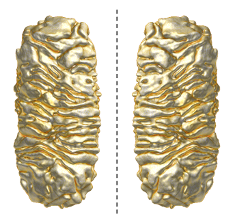
Je kan de twee varianten van de molecule vergelijken met onze handen. Je linker- en rechterhand zijn spiegelbeelden van elkaar, maar er is geen enkele manier om ze exact met elkaar te laten overlappen.
Uiteraard kan je wel het onderscheid maken tussen je linker- en je rechterhand. Je kijkt er gewoon naar. Dat gaat niet bij een molecule. Die is daarvoor gewoonweg te klein. Om te weten met welke molecule we als wetenschappers te maken hebben, trekken we naar het lab. Daar kunnen we meten welke soort molecule in de potjes zit. Maar wat blijkt? Die gespiegelde varianten zitten samen in hetzelfde potje, en we kunnen de twee varianten niet gemakkelijk van elkaar splitsen.

Onze handen zijn elkaars spiegelbeeld, maar er is geen enkele manier om ze exact met elkaar te laten overlappen.
Nanotechnologie to the rescue!
In mijn onderzoek heb ik nanodeeltjes bestudeerd die een mogelijke oplossing kunnen bieden. Dat zijn deeltjes met afmetingen die 1.000 tot wel 100.000 keer kleiner kunnen zijn dan de dikte van een haar. En dat maakt ze heel bijzonder.
De vorm van nanodeeltjes bepaalt namelijk hoe ze zich gedragen. We kunnen nanodeeltjes maken met allerlei exotische vormen en al even exotische eigenschappen. Sinds kort kunnen we zelfs nanodeeltjes maken die ook een spiegelbeeld hebben dat zich anders gedraagt. Die zouden we kunnen gebruiken om de twee varianten van een molecule uit elkaar te halen, wat de productie van betere medicijnen mogelijk maakt.
Sommige nanodeeltjes hebben een spiegelbeeld dat zich anders gedraagt.
Met de juiste vorm kunnen we gespiegelde nanodeeltjes gebruiken als politieagenten. Als we ze toevoegen aan een medicijn waar nog boosaardige moleculen in zitten, dan kunnen die nanodeeltjes deze moleculen opsporen en vastgrijpen. Zo worden de slechte spiegelbeelden van moleculen uitgeschakeld en kunnen we betere medicijnen maken.
Er zijn dan ook veel wetenschappers die hier de laatste jaren hard aan werken. Maar verrassend genoeg is de exacte invloed van de vorm van zulke gespiegelde nanodeeltjes op hun eigenschappen nog niet gekend. Daarom heb ik in mijn eindwerk een nieuwe onderzoeksmethode ontworpen om het onderzoek rond deze nanodeeltjes te vergemakkelijken.
Links of rechts?
Het is niet eenvoudig om de vorm van zulke nanodeeltjes te onderzoeken, want nanodeeltjes zijn zelfs met de beste optische microscoop niet zichtbaar. Ze zijn te klein. Je hebt dus een elektronenmicroscoop nodig, die nog veel sterker kan vergroten. Daarmee kan je dan een foto nemen, waarop je het nanodeeltje langs één kant kan bekijken. Om de volledige vorm van het nanodeeltje te achterhalen, nemen we een reeks foto’s langs verschillende kanten. Door deze te combineren, kunnen we een volledige driedimensionale virtuele kopie van het nanodeeltje berekenen.
Nu willen we graag weten welke van de twee spiegelbeeldvarianten we bekeken hebben. Links of rechts? En daar knelt het schoentje. Die beslissing gebeurt momenteel nog door de subjectieve interpretatie van de onderzoeker, maar dat is niet voldoende als we die nanodeeltjes echt willen begrijpen. Het is ook belangrijk om te weten hoe sterk een nanodeeltje verschilt van zijn spiegelbeeld. Hoe groter het verschil, hoe beter het zal werken.
Meten is weten
Daarom heb ik tijdens mijn onderzoek een methode ontworpen die eenduidig kan bepalen met welke van de twee spiegelbeelden men te maken heeft. Bovendien kan ik bepalen hoe sterk een nanodeeltje van zijn spiegelbeeld verschilt. Dat was tot nu toe nog niet mogelijk. Vervolgens heb ik de handen in elkaar geslagen met een internationaal team van onderzoekers die gespecialiseerd zijn in de productie van nanodeeltjes. Samen hebben we al enkele manieren om de nanodeeltjes te maken in detail onderzocht en geoptimaliseerd met de hulp van deze nieuwe onderzoeksmethode.
Door gebruik te maken van deze nieuwe methode, is het nu mogelijk om de spiegelbeelden van nanodeeltjes op een objectieve manier te onderzoeken. Dat zal ertoe leiden dat we de eigenschappen van nanodeeltjes goed begrijpen. Op haar beurt kunnen we dan nieuwe nanodeeltjes maken waarmee we de boosaardige variant van een molecule kunnen betrappen en uitschakelen. Dit kan onze medicijnen alleen maar verbeteren!
Bibliografie
(1) Mock, J. J.; Hill, R. T.; Degiron, A.; Zauscher, S.; Chilkoti, A.; Smith, D. R. Distance-Dependent Plasmon Resonant Coupling between a Gold Nanoparticle and Gold Film. Nano Lett. 2008, 8 (8), 2245–2252. https://doi.org/10.1021/nl080872f.
(2) Nehl, C. L.; Hafner, J. H. Shape-Dependent Plasmon Resonances of Gold Nanoparticles. Journal of Materials Chemistry. The Royal Society of Chemistry May 13, 2008, pp 2415–2419. https://doi.org/10.1039/b714950f.
(3) Jain, P. K.; Lee, K. S.; El-Sayed, I. H.; El-Sayed, M. A. Calculated Absorption and Scattering Properties of Gold Nanoparticles of Different Size, Shape, and Composition: Applications in Biological Imaging and Biomedicine. J. Phys. Chem. B 2006, 110 (14), 7238–7248. https://doi.org/10.1021/jp057170o.
(4) Yaqoob, A. A.; Ahmad, H.; Parveen, T.; Ahmad, A.; Oves, M.; Ismail, I. M. I.; Qari, H. A.; Umar, K.; Mohamad Ibrahim, M. N. Recent Advances in Metal Decorated Nanomaterials and Their Various Biological Applications: A Review. Frontiers in Chemistry. Frontiers Media S.A. May 19, 2020, p 341. https://doi.org/10.3389/fchem.2020.00341.
(5) Garcia, M. A. Surface Plasmons in Metallic Nanoparticles: Fundamentals and Applications. Journal of Physics D: Applied Physics. IOP Publishing June 24, 2011, p 283001. https://doi.org/10.1088/0022-3727/44/28/283001.
(6) Kumar, J.; Eraña, H.; López-Martínez, E.; Claes, N.; Martín, V. F.; Solís, D. M.; Bals, S.; Cortajarena, A. L.; Castilla, J.; Liz-Marzán, L. M. Detection of Amyloid Fibrils in Parkinson’s Disease Using Plasmonic Chirality. Proc. Natl. Acad. Sci. 2018, 115 (13), 3225–3230. https://doi.org/10.1073/pnas.1721690115.
(7) Jimenez de Aberasturi, D.; Belén Serrano-Montes, A.; Liz-Marzán, L. M.; Liz-Marzán, L. M.; Jimenez de Aberasturi, D.; Serrano-Montes, A. B. Modern Applications of Plasmonic Nanoparticles: From Energy to Health. Adv. Opt. Mater. 2015, 3 (5), 602–617. https://doi.org/10.1002/ADOM.201500053.
(8) Esposito, M.; Tasco, V.; Todisco, F.; Cuscunà, M.; Benedetti, A.; Sanvitto, D.; Passaseo, A. Triple-Helical Nanowires by Tomographic Rotatory Growth for Chiral Photonics. Nat. Commun. 2015, 6 (1), 1–7. https://doi.org/10.1038/ncomms7484.
(9) Frank, B.; Yin, X.; Schäferling, M.; Zhao, J.; Hein, S. M.; Braun, P. V.; Giessen, H. Large-Area 3D Chiral Plasmonic Structures. ACS Nano 2013, 7 (7), 6321–6329. https://doi.org/10.1021/nn402370x.
(10) Furusawa, G.; Kan, T. Au Nanospirals Transferred onto PDMS Film Exhibiting Circular Dichroism at Visiblewavelengths. Micromachines 2020, 11 (7). https://doi.org/10.3390/MI11070641.
(11) Lee, H. E.; Kim, R. M.; Ahn, H. Y.; Lee, Y. Y.; Byun, G. H.; Im, S. W.; Mun, J.; Rho, J.; Nam, K. T. Cysteine-Encoded Chirality Evolution in Plasmonic Rhombic Dodecahedral Gold Nanoparticles. Nat. Commun. 2020, 11 (1), 1–10. https://doi.org/10.1038/s41467-019-14117-x.
(12) Sobczak, K.; Turczyniak-Surdacka, S.; Lewandowski, W.; Baginski, M.; Tupikowska, M.; González-Rubio, G.; Wójcik, M.; Carlsson, A.; Donten, M. STEM Tomography of Au Helical Assemblies. Microsc. Microanal. 2021, 1–5. https://doi.org/10.1017/S1431927621012009.
(13) González-Rubio, G.; Mosquera, J.; Kumar, V.; Pedrazo-Tardajos, A.; Llombart, P.; Solís, D. M.; Lobato, I.; Noya, E. G.; Guerrero-Martínez, A.; Taboada, J. M.; Obelleiro, F.; MacDowell, L. G.; Bals, S.; Liz-Marzán, L. M. Micelle-Directed Chiral Seeded Growth on Anisotropic Gold Nanocrystals. Science (80-. ). 2020, 368 (6498), 1472–1477. https://doi.org/10.1126/science.aba0980.
(14) Liu, Z.; Xu, Y.; Ji, C. Y.; Chen, S.; Li, X.; Zhang, X.; Yao, Y.; Li, J. Fano-Enhanced Circular Dichroism in Deformable Stereo Metasurfaces. Adv. Mater. 2020, 32 (8). https://doi.org/10.1002/adma.201907077.
(15) Chen, J.; Gao, X.; Zheng, Q.; Liu, J.; Meng, D.; Li, H.; Cai, R.; Fan, H.; Ji, Y.; Wu, X. Bottom-Up Synthesis of Helical Plasmonic Nanorods and Their Application in Generating Circularly Polarized Luminescence. ACS Nano 2021, 15 (9), 15114–15122. https://doi.org/10.1021/ACSNANO.1C05489.
(16) Cho, N. H.; Lee, H. E.; Ahn, H. Y.; Lee, Y. Y.; Im, S. W.; Kim, H.; Nam, K. T. Cysteine Induced Chiral Morphology in Palladium Nanoparticle. Part. Part. Syst. Charact. 2019, 36 (5), 1900062. https://doi.org/10.1002/ppsc.201900062.
(17) Cho, N. H.; Byun, G. H.; Lim, Y. C.; Im, S. W.; Kim, H.; Lee, H. E.; Ahn, H. Y.; Nam, K. T. Uniform Chiral Gap Synthesis for High Dissymmetry Factor in Single Plasmonic Gold Nanoparticle. ACS Nano 2020, 14 (3), 3595–3602. https://doi.org/10.1021/acsnano.9b10094.
(18) Lee, H. E.; Ahn, H. Y.; Mun, J.; Lee, Y. Y.; Kim, M.; Cho, N. H.; Chang, K.; Kim, W. S.; Rho, J.; Nam, K. T. Amino-Acid- and Peptide-Directed Synthesis of Chiral Plasmonic Gold Nanoparticles. Nature 2018, 556 (7701), 360–364. https://doi.org/10.1038/s41586-018-0034-1.
(19) Sample Courtesy of Dr. Bing Ni, University of Konstanz.
(20) Hentschel, M.; Schäferling, M.; Duan, X.; Giessen, H.; Liu, N. Chiral Plasmonics. Science Advances. American Association for the Advancement of Science May 1, 2017, p e1602735. https://doi.org/10.1126/sciadv.1602735.
(21) Goerlitzer, E. S. A.; Puri, A. S.; Moses, J. J.; Poulikakos, L. V.; Vogel, N. The Beginner’s Guide to Chiral Plasmonics: Mostly Harmless Theory and the Design of Large-Area Substrates. Advanced Optical Materials. May 28, 2021, p 2100378. https://doi.org/10.1002/adom.202100378.
(22) Gutsche, P.; Garcia-Santiago, X.; Schneider, P. I.; McPeak, K. M.; Nieto-Vesperinas, M.; Burger, S. Role of Geometric Shape in Chiral Optics. Symmetry (Basel). 2020, 12 (1). https://doi.org/10.3390/SYM12010158.
(23) Ben-Moshe, A.; da Silva, A.; Muller, A.; Abu-Odeh, A.; Harrison, P.; Waelder, J.; Niroui, F.; Ophus, C.; Minor, A. M.; Asta, M.; Theis, W.; Ercius, P.; Alivisatos, A. P. The Chain of Chirality Transfer in Tellurium Nanocrystals. Science (80-. ). 2021, 372 (6543), 729–733. https://doi.org/10.1126/science.abf9645.
(24) Canfield, B. K.; Kujala, S.; Laiho, K.; Jefimovs, K.; Turunen, J.; Kauranen, M. Chirality Arising from Small Defects in Gold Nanoparticle Arrays. Opt. Express 2006, 14 (2), 950. https://doi.org/10.1364/opex.14.000950.
(25) Spaeth, P.; Adhikari, S.; Le, L.; Jollans, T.; Pud, S.; Albrecht, W.; Bauer, T.; Caldarola, M.; Kuipers, L.; Orrit, M. Circular Dichroism Measurement of Single Metal Nanoparticles Using Photothermal Imaging. Nano Lett. 2019, 19 (12), 8934–8940. https://doi.org/10.1021/acs.nanolett.9b03853.
(26) Karst, J.; Cho, N. H.; Kim, H.; Lee, H. E.; Nam, K. T.; Giessen, H.; Hentschel, M. Chiral Scatterometry on Chemically Synthesized Single Plasmonic Nanoparticles. ACS Nano 2019, 13 (8), 8659–8668. https://doi.org/10.1021/acsnano.9b04046.
(27) Williams, D. B.; Carter, C. B. Transmission Electron Microscopy: A Textbook for Materials Science; Springer US, 2009. https://doi.org/10.1007/978-0-387-76501-3.
(28) Nellist, P. D. Scanning Transmission Electron Microscopy; Pennycook, S. J., Nellist, P. D., Eds.; Springer, 2019. https://doi.org/10.1007/978-3-030-00069-1_2.
(29) Zu, S.; Bao, Y.; Fang, Z. Planar Plasmonic Chiral Nanostructures. Nanoscale 2016, 8 (7), 3900–3905. https://doi.org/10.1039/c5nr09302c.
(30) Weyland, M.; Midgley, P. A. Electron Tomography. Mater. Today 2004, 7 (12), 32–40. https://doi.org/10.1016/S1369-7021(04)00569-3.
(31) Radon, J. Über Die Bestimmung von Funktionen Durch Ihre Integralwerte Ängs Gewisser Mannigfaltigkeiten. Akad. Wiss. 1917, 69, 262–277.
(32) Andersen, M. S.; Batenburg, K. J.; Dong, Y.; Quinto, E. T.; Sijbers, J.; Hansen, P. C.; Jorgensen, J. S.; Lionheart, W. R. B. Computed Tomography: Algorithms, Insight, and Just Enough Theory; Hansen, P. C., Jørgensen, J., Lionheart, W. R. B., Eds.; Society for Industrial and Applied Mathematics: Philadelphia, PA, 2021. https://doi.org/10.1137/1.9781611976670.
(33) Gregor, J.; Benson, T. Computational Analysis and Improvement of SIRT. IEEE Trans. Med. Imaging 2008, 27 (7), 918–924. https://doi.org/10.1109/TMI.2008.923696.
(34) Levitan, E.; Herman, G. T. A Maximum A Posteriori Probability Expectation Maximization Algorithm for Image Reconstruction in Emission Tomography. IEEE Trans. Med. Imaging 1987, 6 (3), 185–192. https://doi.org/10.1109/TMI.1987.4307826.
(35) Bruyant, P. P. Analytic and Iterative Reconstruction Algorithms in SPECT. J. Nucl. Med. 2002, 43 (10), 1343–1358.
(36) Goris, B.; Van den Broek, W.; Batenburg, K. J.; Heidari Mezerji, H.; Bals, S. Electron Tomography Based on a Total Variation Minimization Reconstruction Technique. Ultramicroscopy 2012, 113, 120–130. https://doi.org/10.1016/J.ULTRAMIC.2011.11.004.
(37) Vanrompay, H.; Skorikov, A.; Bladt, E.; Béché, A.; Freitag, B.; Verbeeck, J.; Bals, S. Fast versus Conventional HAADF-STEM Tomography of Nanoparticles: Advantages and Challenges. Ultramicroscopy 2021, 221, 113191. https://doi.org/10.1016/j.ultramic.2020.113191.
(38) Altantzis, T.; Lobato, I.; De Backer, A.; Béché, A.; Zhang, Y.; Basak, S.; Porcu, M.; Xu, Q.; Sánchez-Iglesias, A.; Liz-Marzán, L. M.; Van Tendeloo, G.; Van Aert, S.; Bals, S. Three-Dimensional Quantification of the Facet Evolution of Pt Nanoparticles in a Variable Gaseous Environment. Nano Lett. 2019, 19 (1), 477–481. https://doi.org/10.1021/acs.nanolett.8b04303.
(39) Weyland, M. Two and Three Dimensional Nanoscale Analysis : New Techniques and Applications, University of Cambridge, 2001. https://doi.org/https://doi.org/10.17863/CAM.19108.
(40) Kelvin, W. T. The Molecular Tactics of a Crystal; 1894.
(41) Silvestri, I. P.; Colbon, P. J. J. The Growing Importance of Chirality in 3D Chemical Space Exploration and Modern Drug Discovery Approaches for Hit-ID. ACS Med. Chem. Lett. 2021, 12 (8), 1220–1229. https://doi.org/10.1021/acsmedchemlett.1c00251.
(42) H. Brooks, W.; C. Guida, W.; G. Daniel, K. The Significance of Chirality in Drug Design and Development. Curr. Top. Med. Chem. 2011, 11 (7), 760–770. https://doi.org/10.2174/156802611795165098.
(43) Yewande, E. O.; Neal, M. P.; Low, R. The Hausdorff Chirality Measure and a Proposed Hausdorff Structure Measure. Mol. Phys. 2009, 107 (3), 281–291. https://doi.org/10.1080/00268970902835611.
(44) Winckelmans, N.; Altantzis, T.; Grzelczak, M.; Sánchez-Iglesias, A.; Liz-Marzán, L. M.; Bals, S. Multimode Electron Tomography as a Tool to Characterize the Internal Structure and Morphology of Gold Nanoparticles. J. Phys. Chem. C 2018, 122 (25), 13522–13528. https://doi.org/10.1021/acs.jpcc.7b12379.
(45) Heyvaert, W.; Pedrazo-Tardajos, A.; Kadu, A.; Claes, N.; González-Rubio, G.; Liz-Marzán, L. M.; Albrecht, W.; Bals, S. Quantification of the Helical Morphology of Chiral Gold Nanorods. ACS Mater. Lett. 2022, 4 (4), 642–649. https://doi.org/10.1021/acsmaterialslett.2c00055.
(46) Milagres de Oliveira, T.; Albrecht, W.; González-Rubio, G.; Altantzis, T.; Lobato Hoyos, I. P.; Béché, A.; Van Aert, S.; Guerrero-Martínez, A.; Liz-Marzán, L. M.; Bals, S. 3D Characterization and Plasmon Mapping of Gold Nanorods Welded by Femtosecond Laser Irradiation. ACS Nano 2020, 14 (10), 12558–12570. https://doi.org/10.1021/acsnano.0c02610.
(47) Otsu, N. A Threshold Selection Method from Gray-Level Histograms. IEEE Trans. Syst. Man. Cybern. 1979, 9 (1), 62–66. https://doi.org/10.1109/TSMC.1979.4310076.
(48) Student. The Probable Error of a Mean. Biometrika 1908, 6 (1), 1. https://doi.org/10.2307/2331554.
(49) Albrecht, W.; Bals, S. Fast Electron Tomography for Nanomaterials. J. Phys. Chem. C 2020, 124 (50), 27276–27286. https://doi.org/10.1021/acs.jpcc.0c08939.
(50) Vanrompay, H.; Bladt, E.; Albrecht, W.; Béché, A.; Zakhozheva, M.; Sánchez-Iglesias, A.; Liz-Marzán, L. M.; Bals, S. 3D Characterization of Heat-Induced Morphological Changes of Au Nanostars by Fast in Situ Electron Tomography. Nanoscale 2018, 10 (48), 22792–22801. https://doi.org/10.1039/C8NR08376B.
(51) Tiwari, M. P.; Prasad, A. Molecularly Imprinted Polymer Based Enantioselective Sensing Devices: A Review. Anal. Chim. Acta 2015, 853 (1), 1–18. https://doi.org/10.1016/j.aca.2014.06.011.
(52) Aboul-Enein, H. Y.; Bounoua, N.; Rebizi, M.; Wagdy, H. Application of Nanoparticles in Chiral Analysis and Chiral Separation. Chirality. John Wiley & Sons, Ltd May 1, 2021, pp 196–208. https://doi.org/10.1002/chir.23303.
(53) Hao, C.; Xu, L.; Kuang, H.; Xu, C. Artificial Chiral Probes and Bioapplications. Adv. Mater. 2020, 32 (41), 1802075. https://doi.org/10.1002/adma.201802075.
(54) Yuan, Z.; Zhou, Y.; Qiao, Z.; Eng Aik, C.; Tu, W. C.; Wu, X.; Chen, Y. C. Stimulated Chiral Light-Matter Interactions in Biological Microlasers. ACS Nano 2021, 15 (5), 8965–8975. https://doi.org/10.1021/acsnano.1c01805.
(55) Albrecht, W.; Arslan Irmak, E.; Altantzis, T.; Pedrazo-Tardajos, A.; Skorikov, A.; Deng, T. S.; van der Hoeven, J. E. S.; van Blaaderen, A.; Van Aert, S.; Bals, S. 3D Atomic-Scale Dynamics of Laser-Light-Induced Restructuring of Nanoparticles Unraveled by Electron Tomography. Adv. Mater. 2021, 33 (33), 2100972. https://doi.org/10.1002/adma.202100972.
(56) Qu, D.; Archimi, M.; Camposeo, A.; Pisignano, D.; Zussman, E. Circularly Polarized Laser with Chiral Nematic Cellulose Nanocrystal Cavity. ACS Nano 2021, 15 (5), 8753–8760. https://doi.org/10.1021/acsnano.1c01001.
(57) González-Rubio, G.; Kumar, V.; Llombart, P.; Díaz-Núñez, P.; Bladt, E.; Altantzis, T.; Bals, S.; Peña-Rodríguez, O.; Noya, E. G.; MacDowell, L. G.; Guerrero-Martínez, A.; Liz-Marzán, L. M. Disconnecting Symmetry Breaking from Seeded Growth for the Reproducible Synthesis of High Quality Gold Nanorods. ACS Nano 2019, 13 (4), 4424–4435. https://doi.org/10.1021/acsnano.8b09658.